 Movie
Movie Controller

[English] 日本語
 Yorodumi
- PDB-7sbd: Murine Fab/IgE in complex with profilin from Hevea brasieliensis ... -
+ Open data

- Basic information
| Entry | Database: PDB / ID: 7sbd | ||||||
|---|---|---|---|---|---|---|---|
| Title | Murine Fab/IgE in complex with profilin from Hevea brasieliensis (Hev b 8) | ||||||
 Components Components |
| ||||||
 Keywords Keywords | ALLERGEN/IMMUNE SYSTEM / Antibody / Allergen / IgE/Fab fragment / complex / ALLERGEN-IMMUNE SYSTEM complex | ||||||
| Function / homology |  Function and homology information Function and homology information | ||||||
| Biological species |   Hevea brasiliensis (rubber tree) Hevea brasiliensis (rubber tree) | ||||||
| Method |  X-RAY DIFFRACTION / SYNCHROTRON / MOLECULAR REPLACEMENT / Resolution: 3.04 Å X-RAY DIFFRACTION / SYNCHROTRON / MOLECULAR REPLACEMENT / Resolution: 3.04 Å | ||||||
 Authors Authors | Rodriguez-Romero, A. / Garcia-Ramirez, B. | ||||||
| Funding support |  Mexico, 1items Mexico, 1items
| ||||||
 Citation Citation | Journal: Commun Biol / Year: 2022 Title: A native IgE in complex with profilin provides insights into allergen recognition and cross-reactivity. Authors: Garcia-Ramirez, B. / Mares-Mejia, I. / Rodriguez-Hernandez, A. / Cano-Sanchez, P. / Torres-Larios, A. / Ortega, E. / Rodriguez-Romero, A. | ||||||
| History |
|
- Structure visualization
| Structure viewer | Molecule: MolmilJmol/JSmol |
|---|
- Downloads & links
-Download
| PDBx/mmCIF format | 7sbd.cif.gz | 145.1 KB | Display | PDBx/mmCIF format |
|---|---|---|---|---|
| PDB format | pdb7sbd.ent.gz | 88.6 KB | Display | PDB format |
| PDBx/mmJSON format | 7sbd.json.gz | Tree view | PDBx/mmJSON format | |
| Others |  Other downloads Other downloads |
-Validation report
| Arichive directory | https://data.pdbj.org/pub/pdb/validation_reports/sb/7sbdftp://data.pdbj.org/pub/pdb/validation_reports/sb/7sbd | HTTPS FTP |
|---|
-Related structure data
| Related structure data | 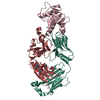 7sbgC 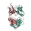 7sd2C 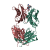 1bafS  5fdsS S: Starting model for refinement C: citing same article ( |
|---|---|
| Similar structure data |
-Links
 PDBj
PDBj


- Assembly
| Deposited unit | 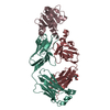
| ||||||||||||
|---|---|---|---|---|---|---|---|---|---|---|---|---|---|
| 1 |
| ||||||||||||
| Unit cell |
|
-Components
| #1: Antibody | Mass: 23226.309 Da / Num. of mol.: 1 / Source method: isolated from a natural source / Source: (natural) |
|---|---|
| #2: Antibody | Mass: 23616.020 Da / Num. of mol.: 1 / Source method: isolated from a natural source / Source: (natural) |
| #3: Protein | Mass: 14636.482 Da / Num. of mol.: 1 Source method: isolated from a genetically manipulated source Source: (gene. exp.) Hevea brasiliensis (rubber tree) / Gene: PRO2 / Production host:  |
| #4: Polysaccharide | alpha-D-mannopyranose-(1-3)-beta-D-mannopyranose-(1-4)-2-acetamido-2-deoxy-beta-D-glucopyranose-(1- ...alpha-D-mannopyranose-(1-3)-beta-D-mannopyranose-(1-4)-2-acetamido-2-deoxy-beta-D-glucopyranose-(1-4)-[alpha-L-fucopyranose-(1-6)]2-acetamido-2-deoxy-beta-D-glucopyranose Source method: isolated from a genetically manipulated source |
| Has ligand of interest | N |
| Has protein modification | Y |
-Experimental details
-Experiment
| Experiment | Method: X-RAY DIFFRACTION / Number of used crystals: 1 |
|---|
- Sample preparation
| Crystal | Density Matthews: 2.49 Å3/Da / Density % sol: 50.62 % |
|---|---|
| Crystal grow | Temperature: 291 K / Method: vapor diffusion, sitting drop / pH: 6.5 Details: 2% Tacsimate, pH 6.0, 0.1 M Bis-Tris, pH 6.5, 20% w/v PEG3350 |
-Data collection
| Diffraction | Mean temperature: 100 K / Serial crystal experiment: N |
|---|---|
| Diffraction source | Source: SYNCHROTRON / Site: NSLS-II  / Beamline: 17-ID-1 / Wavelength: 0.92 Å / Beamline: 17-ID-1 / Wavelength: 0.92 Å |
| Detector | Type: DECTRIS EIGER2 X 9M / Detector: PIXEL / Date: Mar 9, 2018 |
| Radiation | Protocol: SINGLE WAVELENGTH / Monochromatic (M) / Laue (L): M / Scattering type: x-ray |
| Radiation wavelength | Wavelength: 0.92 Å / Relative weight: 1 |
| Reflection | Resolution: 3.04→29.42 Å / Num. obs: 12929 / % possible obs: 99.02 % / Redundancy: 2 % / Biso Wilson estimate: 68.58 Å2 / CC1/2: 0.995 / CC star: 0.999 / Net I/σ(I): 7.12 |
| Reflection shell | Resolution: 3.04→3.152 Å / Rmerge(I) obs: 0.271 / Num. unique obs: 1181 / CC1/2: 0.853 |
- Processing
| Software |
| ||||||||||||||||||||||||||||||||||||||||||||||||||||||||||||||||||||||
|---|---|---|---|---|---|---|---|---|---|---|---|---|---|---|---|---|---|---|---|---|---|---|---|---|---|---|---|---|---|---|---|---|---|---|---|---|---|---|---|---|---|---|---|---|---|---|---|---|---|---|---|---|---|---|---|---|---|---|---|---|---|---|---|---|---|---|---|---|---|---|---|
| Refinement | Method to determine structure: MOLECULAR REPLACEMENT Starting model: PDB entries 1BAF & 5FDS Resolution: 3.04→29.42 Å / SU ML: 0.3831 / Cross valid method: FREE R-VALUE / σ(F): 1.35 / Phase error: 25.7292 Stereochemistry target values: GeoStd + Monomer Library + CDL v1.2
| ||||||||||||||||||||||||||||||||||||||||||||||||||||||||||||||||||||||
| Solvent computation | Shrinkage radii: 0.9 Å / VDW probe radii: 1.11 Å / Solvent model: FLAT BULK SOLVENT MODEL | ||||||||||||||||||||||||||||||||||||||||||||||||||||||||||||||||||||||
| Displacement parameters | Biso mean: 63.99 Å2 | ||||||||||||||||||||||||||||||||||||||||||||||||||||||||||||||||||||||
| Refinement step | Cycle: LAST / Resolution: 3.04→29.42 Å
| ||||||||||||||||||||||||||||||||||||||||||||||||||||||||||||||||||||||
| Refine LS restraints |
| ||||||||||||||||||||||||||||||||||||||||||||||||||||||||||||||||||||||
| LS refinement shell |
|
