 Movie
Movie Controller

[English] 日本語
 Yorodumi
- PDB-7sb2: Structure of the periplasmic domain of GldM from Capnocytophaga c... -
+ Open data

- Basic information
| Entry | Database: PDB / ID: 7sb2 | |||||||||||||||
|---|---|---|---|---|---|---|---|---|---|---|---|---|---|---|---|---|
| Title | Structure of the periplasmic domain of GldM from Capnocytophaga canimorsus | |||||||||||||||
 Components Components | GldM | |||||||||||||||
 Keywords Keywords | MOTOR PROTEIN / type IX secretion system | |||||||||||||||
| Function / homology |  Function and homology information Function and homology information: / : / GldM second domain / GldM third domain / Gliding motility-associated protein GldM / Gliding motility-associated protein GldM, C-terminal / Gliding motility-associated protein GldM, N-terminal / GldM C-terminal domain / GldM N-terminal domain Similarity search - Domain/homology | |||||||||||||||
| Biological species | Capnocytophaga canimorsus | |||||||||||||||
| Method | ELECTRON MICROSCOPY / single particle reconstruction / cryo EM / Resolution: 3.4 Å | |||||||||||||||
 Authors Authors | Hennell James, R. / Deme, J.C. / Lea, S.M. | |||||||||||||||
| Funding support | European Union, 4items
| |||||||||||||||
 Citation Citation | Journal: mBio / Year: 2022 Title: Structures of the Type IX Secretion/Gliding Motility Motor from across the Phylum . Authors: Rory Hennell James / Justin C Deme / Alicia Hunter / Ben C Berks / Susan M Lea /   Abstract: Gliding motility using cell surface adhesins, and export of proteins by the type IX secretion system (T9SS) are two phylum-specific features of the Bacteroidetes. Both of these processes are ...Gliding motility using cell surface adhesins, and export of proteins by the type IX secretion system (T9SS) are two phylum-specific features of the Bacteroidetes. Both of these processes are energized by the GldLM motor complex, which transduces the proton motive force at the inner membrane into mechanical work at the outer membrane. We previously used cryo-electron microscopy to solve the structure of the GldLM motor core from Flavobacterium johnsoniae at 3.9-Å resolution (R. Hennell James, J. C. Deme, A. Kjaer, F. Alcock, et al., Nat Microbiol 6:221-233, 2021, https://dx.doi.org/10.1038/s41564-020-00823-6). Here, we present structures of homologous complexes from a range of pathogenic and environmental species at up to 3.0-Å resolution. These structures show that the architecture of the GldLM motor core is conserved across the phylum, although there are species-specific differences at the N terminus of GldL. The resolution improvements reveal a cage-like structure that ties together the membrane-proximal cytoplasmic region of GldL and influences gliding function. These findings add detail to our structural understanding of bacterial ion-driven motors that drive the T9SS and gliding motility. Many bacteria in the phylum use the type IX secretion system to secrete proteins across their outer membrane. Most of these bacteria can also glide across surfaces using adhesin proteins that are propelled across the cell surface. Both secretion and gliding motility are driven by the GldLM protein complex, which forms a nanoscale electrochemical motor. We used cryo-electron microscopy to study the structure of the GldLM protein complex from different species, including the human pathogens Porphyromonas gingivalis and Capnocytophaga canimorsus. The organization of the motor is conserved across species, but we find species-specific structural differences and resolve motor features at higher resolution. This work improves our understanding of the type IX secretion system, which is a virulence determinant in human and animal diseases. | |||||||||||||||
| History |
|
- Structure visualization
| Structure viewer | Molecule: MolmilJmol/JSmol |
|---|
- Downloads & links
-Download
| PDBx/mmCIF format | 7sb2.cif.gz | 114.9 KB | Display | PDBx/mmCIF format |
|---|---|---|---|---|
| PDB format | pdb7sb2.ent.gz | 89.2 KB | Display | PDB format |
| PDBx/mmJSON format | 7sb2.json.gz | Tree view | PDBx/mmJSON format | |
| Others |  Other downloads Other downloads |
-Validation report
| Arichive directory | https://data.pdbj.org/pub/pdb/validation_reports/sb/7sb2ftp://data.pdbj.org/pub/pdb/validation_reports/sb/7sb2 | HTTPS FTP |
|---|
-Related structure data
| Related structure data |  24961MC  7satC  7sauC  7saxC  7sazC C: citing same article ( M: map data used to model this data |
|---|---|
| Similar structure data |


-Links
 PDBj
PDBj- Assembly
| Deposited unit | 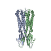
|
|---|---|
| 1 |
|
-Components
| #1: Protein | Mass: 41123.895 Da / Num. of mol.: 2 / Fragment: C-terminal TEV cleavage site and TwinStrep Tag Source method: isolated from a genetically manipulated source Source: (gene. exp.)  Capnocytophaga canimorsus (strain 5) (bacteria) Capnocytophaga canimorsus (strain 5) (bacteria)Gene: Ccan_01630 / Plasmid: pT12 / Production host: Sequence details | C-terminal TEV cleavage site and TwinStrep Tag | |
|---|
-Experimental details
-Experiment
| Experiment | Method: ELECTRON MICROSCOPY |
|---|---|
| EM experiment | Aggregation state: PARTICLE / 3D reconstruction method: single particle reconstruction |
- Sample preparation
| Component | Name: Type IX Secretion System/gliding motility GldM periplasmic domain Type: COMPLEX Details: The structure of the periplasmic domain of GldM was solved using a sample that also included GldL and was used to solve a structure of GldLM Entity ID: all / Source: RECOMBINANT | |||||||||||||||||||||||||
|---|---|---|---|---|---|---|---|---|---|---|---|---|---|---|---|---|---|---|---|---|---|---|---|---|---|---|
| Molecular weight | Experimental value: NO | |||||||||||||||||||||||||
| Source (natural) | Organism: Capnocytophaga canimorsus Cc5 (bacteria) | |||||||||||||||||||||||||
| Source (recombinant) | Organism: | |||||||||||||||||||||||||
| Buffer solution | pH: 8 | |||||||||||||||||||||||||
| Buffer component |
| |||||||||||||||||||||||||
| Specimen | Conc.: 1 mg/ml / Embedding applied: NO / Shadowing applied: NO / Staining applied: NO / Vitrification applied: YES Details: The structure of the periplasmic domain of GldM was solved using a sample that also included GldL and was used to solve a structure of GldLM | |||||||||||||||||||||||||
| Specimen support | Details: 15 mA / Grid material: GOLD / Grid mesh size: 300 divisions/in. / Grid type: Quantifoil R1.2/1.3 | |||||||||||||||||||||||||
| Vitrification | Instrument: FEI VITROBOT MARK IV / Cryogen name: ETHANE / Humidity: 100 % / Chamber temperature: 277.15 K / Details: Wait time 10 s Blot time 2 s |
- Electron microscopy imaging
| Experimental equipment |  Model: Titan Krios / Image courtesy: FEI Company |
|---|---|
| Microscopy | Model: FEI TITAN KRIOS |
| Electron gun | Electron source:  FIELD EMISSION GUN / Accelerating voltage: 300 kV / Illumination mode: FLOOD BEAM FIELD EMISSION GUN / Accelerating voltage: 300 kV / Illumination mode: FLOOD BEAM |
| Electron lens | Mode: BRIGHT FIELD |
| Image recording | Electron dose: 59 e/Å2 / Film or detector model: GATAN K3 BIOQUANTUM (6k x 4k) |
- Processing
| EM software |
| ||||||||||||||||||||||||||||||||
|---|---|---|---|---|---|---|---|---|---|---|---|---|---|---|---|---|---|---|---|---|---|---|---|---|---|---|---|---|---|---|---|---|---|
| CTF correction | Type: PHASE FLIPPING AND AMPLITUDE CORRECTION | ||||||||||||||||||||||||||||||||
| Particle selection | Num. of particles selected: 9197926 | ||||||||||||||||||||||||||||||||
| Symmetry | Point symmetry: C1 (asymmetric) | ||||||||||||||||||||||||||||||||
| 3D reconstruction | Resolution: 3.4 Å / Resolution method: FSC 0.143 CUT-OFF / Num. of particles: 595559 / Symmetry type: POINT | ||||||||||||||||||||||||||||||||
| Atomic model building | B value: 93.02 / Protocol: AB INITIO MODEL / Space: REAL Details: A homology model based on the structure of FjoGldM (PDB 6EY4) was used as a starting model and modified to fit the EM density using Coot. Refinement was carried out using Phenix. |
