 Movie
Movie Controller

[English] 日本語
 Yorodumi
- PDB-7s6k: J08 fragment antigen binding in complex with SARS-CoV-2-6P-Mut2 S... -
+ Open data

- Basic information
| Entry | Database: PDB / ID: 7s6k | |||||||||||||||||||||||||||||||||||||||||||||||||||||||||||||||
|---|---|---|---|---|---|---|---|---|---|---|---|---|---|---|---|---|---|---|---|---|---|---|---|---|---|---|---|---|---|---|---|---|---|---|---|---|---|---|---|---|---|---|---|---|---|---|---|---|---|---|---|---|---|---|---|---|---|---|---|---|---|---|---|---|
| Title | J08 fragment antigen binding in complex with SARS-CoV-2-6P-Mut2 S protein (conformation 2) | |||||||||||||||||||||||||||||||||||||||||||||||||||||||||||||||
 Components Components |
| |||||||||||||||||||||||||||||||||||||||||||||||||||||||||||||||
 Keywords Keywords | VIRAL PROTEIN/Immune System / COVID / SARS / CoV-2 / viral glycoprotein / Spike / stabilizing mutations / coronavirus / ultrapotent antibody / neutralizing antibody / VIRAL PROTEIN / VIRAL PROTEIN-Immune System complex | |||||||||||||||||||||||||||||||||||||||||||||||||||||||||||||||
| Function / homology |  Function and homology information Function and homology informationsymbiont-mediated disruption of host tissue / Maturation of spike protein / Translation of Structural Proteins / Virion Assembly and Release / host cell surface / host extracellular space / viral translation / symbiont-mediated-mediated suppression of host tetherin activity / Induction of Cell-Cell Fusion / structural constituent of virion ...symbiont-mediated disruption of host tissue / Maturation of spike protein / Translation of Structural Proteins / Virion Assembly and Release / host cell surface / host extracellular space / viral translation / symbiont-mediated-mediated suppression of host tetherin activity / Induction of Cell-Cell Fusion / structural constituent of virion / entry receptor-mediated virion attachment to host cell / membrane fusion / Attachment and Entry / host cell endoplasmic reticulum-Golgi intermediate compartment membrane / positive regulation of viral entry into host cell / receptor-mediated virion attachment to host cell / host cell surface receptor binding / symbiont-mediated suppression of host innate immune response / receptor ligand activity / endocytosis involved in viral entry into host cell / fusion of virus membrane with host plasma membrane / fusion of virus membrane with host endosome membrane / viral envelope / symbiont entry into host cell / virion attachment to host cell / SARS-CoV-2 activates/modulates innate and adaptive immune responses / host cell plasma membrane / virion membrane / identical protein binding / membrane / plasma membrane Similarity search - Function | |||||||||||||||||||||||||||||||||||||||||||||||||||||||||||||||
| Biological species |   Severe acute respiratory syndrome coronavirus 2 Severe acute respiratory syndrome coronavirus 2 Homo sapiens (human) Homo sapiens (human) | |||||||||||||||||||||||||||||||||||||||||||||||||||||||||||||||
| Method | ELECTRON MICROSCOPY / single particle reconstruction / cryo EM / Resolution: 3.4 Å | |||||||||||||||||||||||||||||||||||||||||||||||||||||||||||||||
 Authors Authors | Ozorowski, G. / Torres, J.L. / Ward, A.B. | |||||||||||||||||||||||||||||||||||||||||||||||||||||||||||||||
| Funding support |  Italy, 2items Italy, 2items
| |||||||||||||||||||||||||||||||||||||||||||||||||||||||||||||||
 Citation Citation | Journal: Proc Natl Acad Sci U S A / Year: 2022 Title: Structural insights of a highly potent pan-neutralizing SARS-CoV-2 human monoclonal antibody. Authors: Jonathan L Torres / Gabriel Ozorowski / Emanuele Andreano / Hejun Liu / Jeffrey Copps / Giulia Piccini / Lorena Donnici / Matteo Conti / Cyril Planchais / Delphine Planas / Noemi Manganaro / ...Authors: Jonathan L Torres / Gabriel Ozorowski / Emanuele Andreano / Hejun Liu / Jeffrey Copps / Giulia Piccini / Lorena Donnici / Matteo Conti / Cyril Planchais / Delphine Planas / Noemi Manganaro / Elisa Pantano / Ida Paciello / Piero Pileri / Timothée Bruel / Emanuele Montomoli / Hugo Mouquet / Olivier Schwartz / Claudia Sala / Raffaele De Francesco / Ian A Wilson / Rino Rappuoli / Andrew B Ward /   Abstract: As the coronavirus disease 2019 (COVID-19) pandemic continues, there is a strong need for highly potent monoclonal antibodies (mAbs) that are resistant against severe acute respiratory syndrome ...As the coronavirus disease 2019 (COVID-19) pandemic continues, there is a strong need for highly potent monoclonal antibodies (mAbs) that are resistant against severe acute respiratory syndrome coronavirus 2 (SARS-CoV-2) variants of concern (VoCs). Here, we evaluate the potency of the previously described mAb J08 against these variants using cell-based assays and delve into the molecular details of the binding interaction using cryoelectron microscopy (cryo-EM) and X-ray crystallography. We show that mAb J08 has low nanomolar affinity against most VoCs and binds high on the receptor binding domain (RBD) ridge, away from many VoC mutations. These findings further validate the phase II/III human clinical trial underway using mAb J08 as a monoclonal therapy. | |||||||||||||||||||||||||||||||||||||||||||||||||||||||||||||||
| History |
|
- Structure visualization
| Structure viewer | Molecule: MolmilJmol/JSmol |
|---|
- Downloads & links
-Download
| PDBx/mmCIF format | 7s6k.cif.gz | 694.9 KB | Display | PDBx/mmCIF format |
|---|---|---|---|---|
| PDB format | pdb7s6k.ent.gz | 564.8 KB | Display | PDB format |
| PDBx/mmJSON format | 7s6k.json.gz | Tree view | PDBx/mmJSON format | |
| Others |  Other downloads Other downloads |
-Validation report
| Summary document | 7s6k_validation.pdf.gz | 2.1 MB | Display | wwPDB validaton report |
|---|---|---|---|---|
| Full document | 7s6k_full_validation.pdf.gz | 2.1 MB | Display | |
| Data in XML | 7s6k_validation.xml.gz | 112.7 KB | Display | |
| Data in CIF | 7s6k_validation.cif.gz | 178.3 KB | Display | |
| Arichive directory | https://data.pdbj.org/pub/pdb/validation_reports/s6/7s6kftp://data.pdbj.org/pub/pdb/validation_reports/s6/7s6k | HTTPS FTP |
-Related structure data
| Related structure data |  24878MC  7s6iC 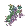 7s6jC 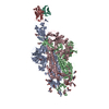 7s6lC 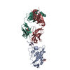 7sbuC M: map data used to model this data C: citing same article ( |
|---|---|
| Similar structure data |


-Links
 PDBj
PDBj


- Assembly
| Deposited unit | 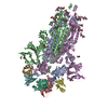
|
|---|---|
| 1 |
|
-Components
| #1: Protein | Mass: 141326.422 Da / Num. of mol.: 3 Mutation: S383C, R682G, R683S, R685S, F817P, A892P, A899P, A942P, D985C, K986P, V987P Source method: isolated from a genetically manipulated source Source: (gene. exp.) Severe acute respiratory syndrome coronavirus 2Gene: S, 2 / Cell line (production host): HEK293F / Production host: Homo sapiens (human) / References: UniProt: P0DTC2#2: Antibody | Mass: 13961.632 Da / Num. of mol.: 3 Source method: isolated from a genetically manipulated source Source: (gene. exp.) Homo sapiens (human) / Cell line (production host): HEK293F / Production host: Homo sapiens (human)#3: Antibody | Mass: 11252.522 Da / Num. of mol.: 3 Source method: isolated from a genetically manipulated source Source: (gene. exp.) Homo sapiens (human) / Cell line (production host): HEK293F / Production host: Homo sapiens (human)#4: Polysaccharide | 2-acetamido-2-deoxy-beta-D-glucopyranose-(1-4)-2-acetamido-2-deoxy-beta-D-glucopyranose Source method: isolated from a genetically manipulated source #5: Sugar | ChemComp-NAG /   Type: D-saccharide, beta linking / Mass: 221.208 Da / Num. of mol.: 21 / Source method: obtained synthetically / Formula: C8H15NO6 Type: D-saccharide, beta linking / Mass: 221.208 Da / Num. of mol.: 21 / Source method: obtained synthetically / Formula: C8H15NO6Has ligand of interest | N | Has protein modification | Y | |
|---|
-Experimental details
-Experiment
| Experiment | Method: ELECTRON MICROSCOPY |
|---|---|
| EM experiment | Aggregation state: PARTICLE / 3D reconstruction method: single particle reconstruction |
- Sample preparation
| Component | Name: J08 fragment antigen binding in complex with SARS-2-CoV-6P-Mut2 S protein Type: COMPLEX / Entity ID: #1-#3 / Source: MULTIPLE SOURCES | ||||||||||||||||||||
|---|---|---|---|---|---|---|---|---|---|---|---|---|---|---|---|---|---|---|---|---|---|
| Molecular weight | Value: 0.69 MDa / Experimental value: NO | ||||||||||||||||||||
| Buffer solution | pH: 8 / Details: Detergent added shortly before freezing | ||||||||||||||||||||
| Buffer component |
| ||||||||||||||||||||
| Specimen | Conc.: 3 mg/ml / Embedding applied: NO / Shadowing applied: NO / Staining applied: NO / Vitrification applied: YES | ||||||||||||||||||||
| Specimen support | Grid material: COPPER / Grid mesh size: 400 divisions/in. / Grid type: Quantifoil R1.2/1.3 | ||||||||||||||||||||
| Vitrification | Instrument: FEI VITROBOT MARK IV / Cryogen name: ETHANE / Humidity: 100 % / Chamber temperature: 277 K / Details: 3 s blot time |
- Electron microscopy imaging
| Experimental equipment |  Model: Talos Arctica / Image courtesy: FEI Company |
|---|---|
| Microscopy | Model: FEI TALOS ARCTICA |
| Electron gun | Electron source:  FIELD EMISSION GUN / Accelerating voltage: 200 kV / Illumination mode: FLOOD BEAM FIELD EMISSION GUN / Accelerating voltage: 200 kV / Illumination mode: FLOOD BEAM |
| Electron lens | Mode: BRIGHT FIELD / Nominal magnification: 36000 X / Nominal defocus max: 2400 nm / Nominal defocus min: 200 nm / Cs: 2.7 mm / Alignment procedure: COMA FREE |
| Specimen holder | Cryogen: NITROGEN / Specimen holder model: FEI TITAN KRIOS AUTOGRID HOLDER |
| Image recording | Average exposure time: 12 sec. / Electron dose: 50 e/Å2 / Detector mode: COUNTING / Film or detector model: GATAN K2 SUMMIT (4k x 4k) |
| Image scans | Movie frames/image: 48 |
- Processing
| Software | Name: PHENIX / Version: 1.19.2_4158: / Classification: refinement | ||||||||||||||||||||||||||||||||||||||||
|---|---|---|---|---|---|---|---|---|---|---|---|---|---|---|---|---|---|---|---|---|---|---|---|---|---|---|---|---|---|---|---|---|---|---|---|---|---|---|---|---|---|
| EM software |
| ||||||||||||||||||||||||||||||||||||||||
| CTF correction | Type: PHASE FLIPPING AND AMPLITUDE CORRECTION | ||||||||||||||||||||||||||||||||||||||||
| Symmetry | Point symmetry: C3 (3 fold cyclic) | ||||||||||||||||||||||||||||||||||||||||
| 3D reconstruction | Resolution: 3.4 Å / Resolution method: FSC 0.143 CUT-OFF / Num. of particles: 52678 / Symmetry type: POINT | ||||||||||||||||||||||||||||||||||||||||
| Atomic model building | Space: REAL | ||||||||||||||||||||||||||||||||||||||||
| Refine LS restraints |
|
