 Movie
Movie Controller


+ Open data

- Basic information
| Entry | Database: PDB / ID: 7s2q | ||||||
|---|---|---|---|---|---|---|---|
| Title | Crystal structure of hen egg white lysozyme | ||||||
 Components Components | Lysozyme C | ||||||
 Keywords Keywords | HYDROLASE | ||||||
| Function / homology |  Function and homology information Function and homology informationLactose synthesis / Antimicrobial peptides / Neutrophil degranulation / beta-N-acetylglucosaminidase activity / cell wall macromolecule catabolic process / lysozyme / lysozyme activity / killing of cells of another organism / defense response to Gram-negative bacterium / defense response to bacterium ...Lactose synthesis / Antimicrobial peptides / Neutrophil degranulation / beta-N-acetylglucosaminidase activity / cell wall macromolecule catabolic process / lysozyme / lysozyme activity / killing of cells of another organism / defense response to Gram-negative bacterium / defense response to bacterium / defense response to Gram-positive bacterium / endoplasmic reticulum / extracellular space / identical protein binding / cytoplasm Similarity search - Function | ||||||
| Biological species |  | ||||||
| Method |  X-RAY DIFFRACTION / MOLECULAR REPLACEMENT / molecular replacement / Resolution: 1.6 Å X-RAY DIFFRACTION / MOLECULAR REPLACEMENT / molecular replacement / Resolution: 1.6 Å | ||||||
 Authors Authors | Lima, L.M.T.R. / Ramos, N.G. | ||||||
| Funding support |  Brazil, 1items Brazil, 1items
| ||||||
 Citation Citation | Journal: Anal.Biochem. / Year: 2022 Title: The reproducible normality of the crystallographic B-factor. Authors: Ramos, N.G. / Sarmanho, G.F. / de Sa Ribeiro, F. / de Souza, V. / Lima, L.M.T.R. | ||||||
| History |
|
- Structure visualization
| Structure viewer | Molecule: MolmilJmol/JSmol |
|---|
- Downloads & links
-Download
| PDBx/mmCIF format | 7s2q.cif.gz | 43.3 KB | Display | PDBx/mmCIF format |
|---|---|---|---|---|
| PDB format | pdb7s2q.ent.gz | 28.2 KB | Display | PDB format |
| PDBx/mmJSON format | 7s2q.json.gz | Tree view | PDBx/mmJSON format | |
| Others |  Other downloads Other downloads |
-Validation report
| Arichive directory | https://data.pdbj.org/pub/pdb/validation_reports/s2/7s2qftp://data.pdbj.org/pub/pdb/validation_reports/s2/7s2q | HTTPS FTP |
|---|
-Related structure data
| Related structure data |  7s27C  7s28C  7s29C  7s2aC  7s2bC 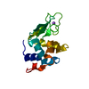 7s2cC  7s2dC  7s2eC  7s2fC  7s2gC  7s2uC  7s2vC 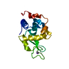 7s2wC  7s30C 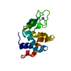 7s31C 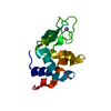 7s32C  7s33C  7s34C  7s35C 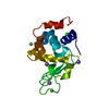 6qwyS S: Starting model for refinement C: citing same article ( |
|---|---|
| Similar structure data |
-Links
 PDBj
PDBj


- Assembly
| Deposited unit | 
| ||||||||||||
|---|---|---|---|---|---|---|---|---|---|---|---|---|---|
| 1 |
| ||||||||||||
| Unit cell |
| ||||||||||||
| Components on special symmetry positions |
|
-Components
| #1: Protein | Mass: 14331.160 Da / Num. of mol.: 1 / Source method: isolated from a natural source / Details: egg white / Source: (natural) |
|---|---|
| #2: Chemical | ChemComp-NA /   Mass: 22.990 Da / Num. of mol.: 1 / Source method: obtained synthetically / Formula: Na Mass: 22.990 Da / Num. of mol.: 1 / Source method: obtained synthetically / Formula: Na |
| #3: Water | ChemComp-HOH /  Mass: 18.015 Da / Num. of mol.: 143 / Source method: isolated from a natural source / Formula: H2O Mass: 18.015 Da / Num. of mol.: 143 / Source method: isolated from a natural source / Formula: H2O |
| Has ligand of interest | N |
| Has protein modification | Y |
-Experimental details
-Experiment
| Experiment | Method: X-RAY DIFFRACTION / Number of used crystals: 1 |
|---|
- Sample preparation
| Crystal | Density Matthews: 1.98 Å3/Da / Density % sol: 37.94 % / Mosaicity: 0.98 ° |
|---|---|
| Crystal grow | Temperature: 295 K / Method: evaporation / pH: 4.6 Details: 1.2 M NaCl, 100 mM Sodium Acetate, 50 mg/mL Lyzozyme, 30% Glycerol for cryoprotection |
-Data collection
| Diffraction | Mean temperature: 125 K / Serial crystal experiment: N | ||||||||||||||||||||||||
|---|---|---|---|---|---|---|---|---|---|---|---|---|---|---|---|---|---|---|---|---|---|---|---|---|---|
| Diffraction source | Source: SEALED TUBE / Type: OXFORD DIFFRACTION ENHANCE ULTRA / Wavelength: 1.54056 Å | ||||||||||||||||||||||||
| Detector | Type: OXFORD TITAN CCD / Detector: CCD / Date: Dec 12, 2020 | ||||||||||||||||||||||||
| Radiation | Monochromator: Ni FILTER / Protocol: SINGLE WAVELENGTH / Monochromatic (M) / Laue (L): M / Scattering type: x-ray | ||||||||||||||||||||||||
| Radiation wavelength | Wavelength: 1.54056 Å / Relative weight: 1 | ||||||||||||||||||||||||
| Reflection | Resolution: 1.6→13.54 Å / Num. obs: 15699 / % possible obs: 99.7 % / Redundancy: 4.5 % / CC1/2: 0.998 / Rmerge(I) obs: 0.06 / Net I/σ(I): 15 / Num. measured all: 71019 / Scaling rejects: 261 | ||||||||||||||||||||||||
| Reflection shell | Diffraction-ID: 1
|
-Phasing
| Phasing | Method: molecular replacement |
|---|
- Processing
| Software |
| ||||||||||||||||||||||||||||||||||||||||||||||||||||||||||||
|---|---|---|---|---|---|---|---|---|---|---|---|---|---|---|---|---|---|---|---|---|---|---|---|---|---|---|---|---|---|---|---|---|---|---|---|---|---|---|---|---|---|---|---|---|---|---|---|---|---|---|---|---|---|---|---|---|---|---|---|---|---|
| Refinement | Method to determine structure: MOLECULAR REPLACEMENT Starting model: 6qwy Resolution: 1.6→13.54 Å / Cor.coef. Fo:Fc: 0.957 / Cor.coef. Fo:Fc free: 0.95 / WRfactor Rfree: 0.2191 / WRfactor Rwork: 0.2057 / FOM work R set: 0.8076 / SU B: 2.2 / SU ML: 0.074 / SU R Cruickshank DPI: 0.1021 / SU Rfree: 0.1077 / Cross valid method: THROUGHOUT / σ(F): 0 / ESU R: 0.102 / ESU R Free: 0.097 / Stereochemistry target values: MAXIMUM LIKELIHOOD Details: isomorphous replacement with REFMAC , restrained refinement with REFMAC, real space refinement with C.O.O.T.
| ||||||||||||||||||||||||||||||||||||||||||||||||||||||||||||
| Solvent computation | Ion probe radii: 0.8 Å / Shrinkage radii: 0.8 Å / VDW probe radii: 1.2 Å / Solvent model: MASK | ||||||||||||||||||||||||||||||||||||||||||||||||||||||||||||
| Displacement parameters | Biso max: 50.84 Å2 / Biso mean: 16.651 Å2 / Biso min: 5.45 Å2
| ||||||||||||||||||||||||||||||||||||||||||||||||||||||||||||
| Refinement step | Cycle: final / Resolution: 1.6→13.54 Å
| ||||||||||||||||||||||||||||||||||||||||||||||||||||||||||||
| Refine LS restraints |
| ||||||||||||||||||||||||||||||||||||||||||||||||||||||||||||
| LS refinement shell | Resolution: 1.6→1.642 Å / Rfactor Rfree error: 0 / Total num. of bins used: 20
|
