 Movie
Movie Controller


+ Open data

- Basic information
| Entry | Database: PDB / ID: 7s0k | ||||||
|---|---|---|---|---|---|---|---|
| Title | HAP2 from Cyanidioschyzon merolae | ||||||
 Components Components | HAP2-GCS1 domain-containing protein | ||||||
 Keywords Keywords | MEMBRANE PROTEIN / Gamete fusogen / Class II fusion protein | ||||||
| Function / homology | Generative cell specific-1/HAP2 domain / Male gamete fusion factor / HAP2/GCS1 / single fertilization / lipid binding / plasma membrane / DI(HYDROXYETHYL)ETHER / Generative cell specific-1/HAP2 domain-containing protein Function and homology information Function and homology information | ||||||
| Biological species |  Cyanidioschyzon merolae (eukaryote) Cyanidioschyzon merolae (eukaryote) | ||||||
| Method |  X-RAY DIFFRACTION / SYNCHROTRON / MOLECULAR REPLACEMENT / Resolution: 2.3 Å X-RAY DIFFRACTION / SYNCHROTRON / MOLECULAR REPLACEMENT / Resolution: 2.3 Å | ||||||
 Authors Authors | Feng, J. / Dong, X.C. / Springer, T.A. | ||||||
| Funding support |  United States, 1items United States, 1items
| ||||||
 Citation Citation | Journal: Nat Commun / Year: 2022 Title: Monomeric prefusion structure of an extremophile gamete fusogen and stepwise formation of the postfusion trimeric state. Authors: Feng, J. / Dong, X. / Su, Y. / Lu, C. / Springer, T.A. | ||||||
| History |
|
- Structure visualization
| Structure viewer | Molecule: MolmilJmol/JSmol |
|---|
- Downloads & links
-Download
| PDBx/mmCIF format | 7s0k.cif.gz | 212.1 KB | Display | PDBx/mmCIF format |
|---|---|---|---|---|
| PDB format | pdb7s0k.ent.gz | 166.5 KB | Display | PDB format |
| PDBx/mmJSON format | 7s0k.json.gz | Tree view | PDBx/mmJSON format | |
| Others |  Other downloads Other downloads |
-Validation report
| Arichive directory | https://data.pdbj.org/pub/pdb/validation_reports/s0/7s0kftp://data.pdbj.org/pub/pdb/validation_reports/s0/7s0k | HTTPS FTP |
|---|
-Related structure data
| Related structure data |  6dbsS S: Starting model for refinement |
|---|---|
| Similar structure data |
-Links
 PDBj
PDBj- Assembly
| Deposited unit | 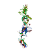
| ||||||||
|---|---|---|---|---|---|---|---|---|---|
| 1 |
| ||||||||
| Unit cell |
|
-Components
-Protein , 1 types, 1 molecules A
| #1: Protein | Mass: 59431.246 Da / Num. of mol.: 1 Source method: isolated from a genetically manipulated source Source: (gene. exp.) Cyanidioschyzon merolae (eukaryote) / Gene: CYME_CMK076C / Production host:  |
|---|
-Sugars , 2 types, 2 molecules 
| #2: Polysaccharide | alpha-D-mannopyranose-(1-3)-beta-D-mannopyranose-(1-3)-beta-D-mannopyranose-(1-4)-2-acetamido-2- ...alpha-D-mannopyranose-(1-3)-beta-D-mannopyranose-(1-3)-beta-D-mannopyranose-(1-4)-2-acetamido-2-deoxy-beta-D-glucopyranose-(1-4)-2-acetamido-2-deoxy-beta-D-glucopyranose Source method: isolated from a genetically manipulated source |
|---|---|
| #3: Sugar | ChemComp-NAG /  Type: D-saccharide, beta linking / Mass: 221.208 Da / Num. of mol.: 1 / Source method: obtained synthetically / Formula: C8H15NO6 Type: D-saccharide, beta linking / Mass: 221.208 Da / Num. of mol.: 1 / Source method: obtained synthetically / Formula: C8H15NO6 |
-Non-polymers , 3 types, 78 molecules 


| #4: Chemical |  Mass: 92.094 Da / Num. of mol.: 3 / Source method: obtained synthetically / Formula: C3H8O3 Mass: 92.094 Da / Num. of mol.: 3 / Source method: obtained synthetically / Formula: C3H8O3#5: Chemical |  Mass: 106.120 Da / Num. of mol.: 2 / Source method: obtained synthetically / Formula: C4H10O3 Mass: 106.120 Da / Num. of mol.: 2 / Source method: obtained synthetically / Formula: C4H10O3#6: Water | ChemComp-HOH / | Mass: 18.015 Da / Num. of mol.: 73 / Source method: isolated from a natural source / Formula: H2O |
|---|
-Details
| Has ligand of interest | N |
|---|---|
| Has protein modification | Y |
-Experimental details
-Experiment
| Experiment | Method: X-RAY DIFFRACTION / Number of used crystals: 1 |
|---|
- Sample preparation
| Crystal | Density Matthews: 3.13 Å3/Da / Density % sol: 60.73 % |
|---|---|
| Crystal grow | Temperature: 293.15 K / Method: vapor diffusion, hanging drop / pH: 6.4 / Details: PEG3350, isopropanol, glycerol, sodium citrate / PH range: 6.2-6.6 |
-Data collection
| Diffraction | Mean temperature: 100 K / Ambient temp details: liquid nitrogen / Serial crystal experiment: N |
|---|---|
| Diffraction source | Source: SYNCHROTRON / Site: APS / Beamline: 23-ID-B / Wavelength: 1.0331 Å |
| Detector | Type: MAR CCD 130 mm / Detector: CCD / Date: Jul 19, 2016 |
| Radiation | Protocol: SINGLE WAVELENGTH / Monochromatic (M) / Laue (L): M / Scattering type: x-ray |
| Radiation wavelength | Wavelength: 1.0331 Å / Relative weight: 1 |
| Reflection | Resolution: 2.3→45.63 Å / Num. obs: 33534 / % possible obs: 98.8 % / Redundancy: 3.3 % / CC1/2: 0.993 / Net I/σ(I): 7.11 |
| Reflection shell | Resolution: 2.3→2.36 Å / Redundancy: 3.2 % / Rmerge(I) obs: 0.2426 / Mean I/σ(I) obs: 0.45 / Num. unique obs: 2329 / CC1/2: 0.225 / % possible all: 94.5 |
- Processing
| Software |
| |||||||||||||||||||||||||||||||||||||||||||||||||||||||||||||||||||||||||||||||||||||||||||
|---|---|---|---|---|---|---|---|---|---|---|---|---|---|---|---|---|---|---|---|---|---|---|---|---|---|---|---|---|---|---|---|---|---|---|---|---|---|---|---|---|---|---|---|---|---|---|---|---|---|---|---|---|---|---|---|---|---|---|---|---|---|---|---|---|---|---|---|---|---|---|---|---|---|---|---|---|---|---|---|---|---|---|---|---|---|---|---|---|---|---|---|---|
| Refinement | Method to determine structure: MOLECULAR REPLACEMENT Starting model: PDB ENTRY 6DBS Resolution: 2.3→45.63 Å / SU ML: 0.49 / Cross valid method: THROUGHOUT / σ(F): 1.33 / Phase error: 34.72 / Stereochemistry target values: ML
| |||||||||||||||||||||||||||||||||||||||||||||||||||||||||||||||||||||||||||||||||||||||||||
| Solvent computation | Shrinkage radii: 0.9 Å / VDW probe radii: 1.11 Å / Solvent model: FLAT BULK SOLVENT MODEL | |||||||||||||||||||||||||||||||||||||||||||||||||||||||||||||||||||||||||||||||||||||||||||
| Displacement parameters | Biso mean: 90.79 Å2 | |||||||||||||||||||||||||||||||||||||||||||||||||||||||||||||||||||||||||||||||||||||||||||
| Refinement step | Cycle: LAST / Resolution: 2.3→45.63 Å
| |||||||||||||||||||||||||||||||||||||||||||||||||||||||||||||||||||||||||||||||||||||||||||
| LS refinement shell |
| |||||||||||||||||||||||||||||||||||||||||||||||||||||||||||||||||||||||||||||||||||||||||||
| Refinement TLS params. | Method: refined / Refine-ID: X-RAY DIFFRACTION
| |||||||||||||||||||||||||||||||||||||||||||||||||||||||||||||||||||||||||||||||||||||||||||
| Refinement TLS group |
|
