 Movie
Movie Controller

[English] 日本語
 Yorodumi
- PDB-7rzz: Crystal structure of FBF-2 in complex with LST-1 site A peptide a... -
+ Open data

- Basic information
| Entry | Database: PDB / ID: 7rzz | ||||||
|---|---|---|---|---|---|---|---|
| Title | Crystal structure of FBF-2 in complex with LST-1 site A peptide and compact FBE RNA | ||||||
 Components Components |
| ||||||
 Keywords Keywords | RNA BINDING PROTEIN / Pumilio RNA-binding proteins | ||||||
| Function / homology |  Function and homology information Function and homology informationsex differentiation / P granule / post-transcriptional regulation of gene expression / G-protein alpha-subunit binding / mRNA 3'-UTR binding / regulation of gene expression / cell differentiation / negative regulation of translation / RNA binding / nucleus / cytoplasm Similarity search - Function | ||||||
| Biological species |  | ||||||
| Method |  X-RAY DIFFRACTION / SYNCHROTRON / MOLECULAR REPLACEMENT / Resolution: 2.39 Å X-RAY DIFFRACTION / SYNCHROTRON / MOLECULAR REPLACEMENT / Resolution: 2.39 Å | ||||||
 Authors Authors | Qiu, C. / Hall, T.M.T. | ||||||
| Funding support |  United States, 1items United States, 1items
| ||||||
 Citation Citation | Journal: Nucleic Acids Res. / Year: 2022 Title: Bipartite interaction sites differentially modulate RNA-binding affinity of a protein complex essential for germline stem cell self-renewal. Authors: Qiu, C. / Wine, R.N. / Campbell, Z.T. / Hall, T.M.T. | ||||||
| History |
|
- Structure visualization
| Structure viewer | Molecule: MolmilJmol/JSmol |
|---|
- Downloads & links
-Download
| PDBx/mmCIF format | 7rzz.cif.gz | 228.5 KB | Display | PDBx/mmCIF format |
|---|---|---|---|---|
| PDB format | pdb7rzz.ent.gz | 149.1 KB | Display | PDB format |
| PDBx/mmJSON format | 7rzz.json.gz | Tree view | PDBx/mmJSON format | |
| Others |  Other downloads Other downloads |
-Validation report
| Summary document | 7rzz_validation.pdf.gz | 447.6 KB | Display | wwPDB validaton report |
|---|---|---|---|---|
| Full document | 7rzz_full_validation.pdf.gz | 449.6 KB | Display | |
| Data in XML | 7rzz_validation.xml.gz | 16.7 KB | Display | |
| Data in CIF | 7rzz_validation.cif.gz | 23.3 KB | Display | |
| Arichive directory | https://data.pdbj.org/pub/pdb/validation_reports/rz/7rzzftp://data.pdbj.org/pub/pdb/validation_reports/rz/7rzz | HTTPS FTP |
-Related structure data
| Related structure data |  7s02C  3k5qS S: Starting model for refinement C: citing same article ( |
|---|---|
| Similar structure data |
-Links
 PDBj
PDBj- Assembly
| Deposited unit | 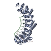
| ||||||||||||
|---|---|---|---|---|---|---|---|---|---|---|---|---|---|
| 1 |
| ||||||||||||
| Unit cell |
|
-Components
| #1: Protein | Mass: 47019.055 Da / Num. of mol.: 1 / Fragment: UNP residues 164-575 Source method: isolated from a genetically manipulated source Source: (gene. exp.)  |
|---|---|
| #2: RNA chain | Mass: 2872.750 Da / Num. of mol.: 1 / Source method: obtained synthetically / Source: (synth.) |
| #3: Protein/peptide | Mass: 3979.517 Da / Num. of mol.: 1 Source method: isolated from a genetically manipulated source Source: (gene. exp.) |
| #4: Water | ChemComp-HOH /  Mass: 18.015 Da / Num. of mol.: 95 / Source method: isolated from a natural source / Formula: H2O Mass: 18.015 Da / Num. of mol.: 95 / Source method: isolated from a natural source / Formula: H2O |
-Experimental details
-Experiment
| Experiment | Method: X-RAY DIFFRACTION / Number of used crystals: 1 |
|---|
- Sample preparation
| Crystal | Density Matthews: 2.59 Å3/Da / Density % sol: 52.47 % |
|---|---|
| Crystal grow | Temperature: 293 K / Method: vapor diffusion, hanging drop / Details: 30% (v/v) PEG 400, 0.1 M CHES, pH 9.5 |
-Data collection
| Diffraction | Mean temperature: 100 K / Serial crystal experiment: N |
|---|---|
| Diffraction source | Source: SYNCHROTRON / Site: APS / Beamline: 22-ID / Wavelength: 1 Å |
| Detector | Type: DECTRIS EIGER X 16M / Detector: PIXEL / Date: Jun 29, 2019 |
| Radiation | Protocol: SINGLE WAVELENGTH / Monochromatic (M) / Laue (L): M / Scattering type: x-ray |
| Radiation wavelength | Wavelength: 1 Å / Relative weight: 1 |
| Reflection | Resolution: 2.39→35.7 Å / Num. obs: 26102 / % possible obs: 99.5 % / Redundancy: 5.1 % / Biso Wilson estimate: 42.65 Å2 / Rpim(I) all: 0.031 / Net I/σ(I): 14.5 |
| Reflection shell | Resolution: 2.39→2.48 Å / Num. unique obs: 2118 / Rpim(I) all: 0.186 / % possible all: 99.4 |
- Processing
| Software |
| |||||||||||||||||||||||||||||||||||||||||||||||||||||||||||||||||||||||||||||||||||||||||||||||||||||||||||||||||||||||||||||
|---|---|---|---|---|---|---|---|---|---|---|---|---|---|---|---|---|---|---|---|---|---|---|---|---|---|---|---|---|---|---|---|---|---|---|---|---|---|---|---|---|---|---|---|---|---|---|---|---|---|---|---|---|---|---|---|---|---|---|---|---|---|---|---|---|---|---|---|---|---|---|---|---|---|---|---|---|---|---|---|---|---|---|---|---|---|---|---|---|---|---|---|---|---|---|---|---|---|---|---|---|---|---|---|---|---|---|---|---|---|---|---|---|---|---|---|---|---|---|---|---|---|---|---|---|---|---|
| Refinement | Method to determine structure: MOLECULAR REPLACEMENT Starting model: 3k5q Resolution: 2.39→35.7 Å / SU ML: 0.2743 / Cross valid method: FREE R-VALUE / σ(F): 1.39 / Phase error: 22.4784 Stereochemistry target values: GeoStd + Monomer Library + CDL v1.2
| |||||||||||||||||||||||||||||||||||||||||||||||||||||||||||||||||||||||||||||||||||||||||||||||||||||||||||||||||||||||||||||
| Solvent computation | Shrinkage radii: 0.9 Å / VDW probe radii: 1.11 Å / Solvent model: FLAT BULK SOLVENT MODEL | |||||||||||||||||||||||||||||||||||||||||||||||||||||||||||||||||||||||||||||||||||||||||||||||||||||||||||||||||||||||||||||
| Displacement parameters | Biso mean: 50.93 Å2 | |||||||||||||||||||||||||||||||||||||||||||||||||||||||||||||||||||||||||||||||||||||||||||||||||||||||||||||||||||||||||||||
| Refinement step | Cycle: LAST / Resolution: 2.39→35.7 Å
| |||||||||||||||||||||||||||||||||||||||||||||||||||||||||||||||||||||||||||||||||||||||||||||||||||||||||||||||||||||||||||||
| Refine LS restraints |
| |||||||||||||||||||||||||||||||||||||||||||||||||||||||||||||||||||||||||||||||||||||||||||||||||||||||||||||||||||||||||||||
| LS refinement shell |
| |||||||||||||||||||||||||||||||||||||||||||||||||||||||||||||||||||||||||||||||||||||||||||||||||||||||||||||||||||||||||||||
| Refinement TLS params. | Method: refined / Refine-ID: X-RAY DIFFRACTION
| |||||||||||||||||||||||||||||||||||||||||||||||||||||||||||||||||||||||||||||||||||||||||||||||||||||||||||||||||||||||||||||
| Refinement TLS group | Refine-ID: X-RAY DIFFRACTION
|
