 Movie
Movie Controller

[English] 日本語
 Yorodumi
- PDB-7rzx: Crystal Structure of Ferritin grown by microbatch method in prese... -
+ Open data

- Basic information
| Entry | Database: PDB / ID: 7rzx | ||||||
|---|---|---|---|---|---|---|---|
| Title | Crystal Structure of Ferritin grown by microbatch method in presence of agarose and electric field 4.3KV | ||||||
 Components Components | Ferritin light chain | ||||||
 Keywords Keywords | METAL BINDING PROTEIN / ferritin / crystallization / agarose / electric field / metal ions | ||||||
| Function / homology |  Function and homology information Function and homology informationferritin complex / autolysosome / ferric iron binding / autophagosome / iron ion transport / ferrous iron binding / cytoplasmic vesicle / intracellular iron ion homeostasis / iron ion binding / cytoplasm Similarity search - Function | ||||||
| Biological species |  | ||||||
| Method |  X-RAY DIFFRACTION / SYNCHROTRON / MOLECULAR REPLACEMENT / Resolution: 2.44 Å X-RAY DIFFRACTION / SYNCHROTRON / MOLECULAR REPLACEMENT / Resolution: 2.44 Å | ||||||
 Authors Authors | Aditya, S. / Priyadharshine, R. / Maham, I. / Miller, J.D. / Stojanoff, V. | ||||||
| Funding support |  United States, 1items United States, 1items
| ||||||
 Citation Citation | Journal: To Be Published Title: Crystal Structure of Ferritin grown by microbatch method in presence of agarose and electric field 4.3KV Authors: Aditya, S. / Priyadharshine, R. / Maham, I. / Miller, J.D. / Stojanoff, V. | ||||||
| History |
|
- Structure visualization
| Structure viewer | Molecule: MolmilJmol/JSmol |
|---|
- Downloads & links
-Download
| PDBx/mmCIF format | 7rzx.cif.gz | 51.2 KB | Display | PDBx/mmCIF format |
|---|---|---|---|---|
| PDB format | pdb7rzx.ent.gz | 36.2 KB | Display | PDB format |
| PDBx/mmJSON format | 7rzx.json.gz | Tree view | PDBx/mmJSON format | |
| Others |  Other downloads Other downloads |
-Validation report
| Arichive directory | https://data.pdbj.org/pub/pdb/validation_reports/rz/7rzxftp://data.pdbj.org/pub/pdb/validation_reports/rz/7rzx | HTTPS FTP |
|---|
-Related structure data
| Related structure data | 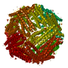 2w0oS S: Starting model for refinement |
|---|---|
| Similar structure data |
-Links
 PDBj
PDBj


- Assembly
| Deposited unit | 
| |||||||||
|---|---|---|---|---|---|---|---|---|---|---|
| 1 | x 24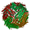
| |||||||||
| Unit cell |
| |||||||||
| Components on special symmetry positions |
|
-Components
| #1: Protein | Mass: 19872.428 Da / Num. of mol.: 1 / Fragment: residue 3-172 Source method: isolated from a genetically manipulated source Source: (gene. exp.)  Escherichia phage EcSzw-2 (virus) / References: UniProt: P02791 Escherichia phage EcSzw-2 (virus) / References: UniProt: P02791 | ||||||
|---|---|---|---|---|---|---|---|
| #2: Chemical | ChemComp-CD /   Mass: 112.411 Da / Num. of mol.: 13 / Source method: obtained synthetically / Formula: Cd Mass: 112.411 Da / Num. of mol.: 13 / Source method: obtained synthetically / Formula: Cd#3: Chemical | ChemComp-SO4 / |   Mass: 96.063 Da / Num. of mol.: 1 / Source method: obtained synthetically / Formula: SO4 Mass: 96.063 Da / Num. of mol.: 1 / Source method: obtained synthetically / Formula: SO4#4: Water | ChemComp-HOH / |  Mass: 18.015 Da / Num. of mol.: 64 / Source method: isolated from a natural source / Formula: H2O Mass: 18.015 Da / Num. of mol.: 64 / Source method: isolated from a natural source / Formula: H2OHas ligand of interest | N | |
-Experimental details
-Experiment
| Experiment | Method: X-RAY DIFFRACTION / Number of used crystals: 1 |
|---|
- Sample preparation
| Crystal | Density Matthews: 3.11 Å3/Da / Density % sol: 60.48 % |
|---|---|
| Crystal grow | Temperature: 290 K / Method: microbatch / Details: CdSO4,NH4SO4,Tris pH7.4 |
-Data collection
| Diffraction | Mean temperature: 100 K / Serial crystal experiment: N |
|---|---|
| Diffraction source | Source: SYNCHROTRON / Site: NSLS-II / Beamline: 19-ID / Wavelength: 0.979 Å |
| Detector | Type: ADSC HF-4M / Detector: PIXEL / Date: Aug 2, 2018 |
| Radiation | Protocol: SINGLE WAVELENGTH / Monochromatic (M) / Laue (L): M / Scattering type: x-ray |
| Radiation wavelength | Wavelength: 0.979 Å / Relative weight: 1 |
| Reflection | Resolution: 2.44→28.63 Å / Num. obs: 9949 / % possible obs: 99.8 % / Redundancy: 20 % / Biso Wilson estimate: 35.88 Å2 / CC1/2: 0.998 / Net I/σ(I): 0.828 |
| Reflection shell | Resolution: 2.44→2.51 Å / Num. unique obs: 704 / CC1/2: 0.766 |
- Processing
| Software |
| ||||||||||||||||||||||||||||||
|---|---|---|---|---|---|---|---|---|---|---|---|---|---|---|---|---|---|---|---|---|---|---|---|---|---|---|---|---|---|---|---|
| Refinement | Method to determine structure: MOLECULAR REPLACEMENT Starting model: 2W0O Resolution: 2.44→28.63 Å / SU ML: 0.26 / Cross valid method: THROUGHOUT / σ(F): 1.36 / Phase error: 22.83 / Stereochemistry target values: ML
| ||||||||||||||||||||||||||||||
| Solvent computation | Shrinkage radii: 0.9 Å / VDW probe radii: 1.11 Å / Solvent model: FLAT BULK SOLVENT MODEL | ||||||||||||||||||||||||||||||
| Displacement parameters | Biso max: 227.7 Å2 / Biso mean: 38.6369 Å2 / Biso min: 22.04 Å2 | ||||||||||||||||||||||||||||||
| Refinement step | Cycle: final / Resolution: 2.44→28.63 Å
| ||||||||||||||||||||||||||||||
| Refine LS restraints |
| ||||||||||||||||||||||||||||||
| LS refinement shell | Refine-ID: X-RAY DIFFRACTION / Rfactor Rfree error: 0 / Total num. of bins used: 4 / % reflection obs: 100 %
|
