 Movie
Movie Controller


+ Open data

- Basic information
| Entry | Database: PDB / ID: 7rw6 | |||||||||||||||||||||||||||
|---|---|---|---|---|---|---|---|---|---|---|---|---|---|---|---|---|---|---|---|---|---|---|---|---|---|---|---|---|
| Title | BORF2-APOBEC3Bctd Complex | |||||||||||||||||||||||||||
 Components Components |
| |||||||||||||||||||||||||||
 Keywords Keywords | OXIDOREDUCTASE/HYDROLASE / Host-pathogen complex / viral protein / antiviral protein / deaminase / OXIDOREDUCTASE-HYDROLASE complex | |||||||||||||||||||||||||||
| Function / homology |  Function and homology information Function and homology informationmRNA Editing: C to U Conversion / Formation of the Editosome / release from viral latency / single-stranded DNA cytosine deaminase / negative regulation of single stranded viral RNA replication via double stranded DNA intermediate / DNA cytosine deamination / cytidine to uridine editing / clearance of foreign intracellular DNA / cytidine deaminase activity / transposable element silencing ...mRNA Editing: C to U Conversion / Formation of the Editosome / release from viral latency / single-stranded DNA cytosine deaminase / negative regulation of single stranded viral RNA replication via double stranded DNA intermediate / DNA cytosine deamination / cytidine to uridine editing / clearance of foreign intracellular DNA / cytidine deaminase activity / transposable element silencing / ribonucleoside-diphosphate reductase / ribonucleoside-diphosphate reductase activity, thioredoxin disulfide as acceptor / deoxyribonucleotide biosynthetic process / detection of maltose stimulus / maltose transport complex / carbohydrate transport / carbohydrate transmembrane transporter activity / maltose binding / maltose transport / maltodextrin transmembrane transport / ATP-binding cassette (ABC) transporter complex, substrate-binding subunit-containing / ATP-binding cassette (ABC) transporter complex / cell chemotaxis / P-body / outer membrane-bounded periplasmic space / defense response to virus / periplasmic space / DNA replication / innate immune response / DNA damage response / RNA binding / zinc ion binding / nucleoplasm / ATP binding / nucleus / membrane / cytoplasm Similarity search - Function | |||||||||||||||||||||||||||
| Biological species |   Human gammaherpesvirus 4 (Epstein-Barr virus) Human gammaherpesvirus 4 (Epstein-Barr virus) Homo sapiens (human) Homo sapiens (human) | |||||||||||||||||||||||||||
| Method | ELECTRON MICROSCOPY / single particle reconstruction / cryo EM / Resolution: 2.55 Å | |||||||||||||||||||||||||||
 Authors Authors | Shaban, N.M. / Yan, R. / Shi, K. / McLellan, J.S. / Yu, Z. / Harris, R.S. | |||||||||||||||||||||||||||
| Funding support |  United States, 1items United States, 1items
| |||||||||||||||||||||||||||
 Citation Citation | Journal: Sci Adv / Year: 2022 Title: Cryo-EM structure of the EBV ribonucleotide reductase BORF2 and mechanism of APOBEC3B inhibition. Authors: Nadine M Shaban / Rui Yan / Ke Shi / Sofia N Moraes / Adam Z Cheng / Michael A Carpenter / Jason S McLellan / Zhiheng Yu / Reuben S Harris / Abstract: Viruses use a plethora of mechanisms to evade immune responses. A recent example is neutralization of the nuclear DNA cytosine deaminase APOBEC3B by the Epstein-Barr virus (EBV) ribonucleotide ...Viruses use a plethora of mechanisms to evade immune responses. A recent example is neutralization of the nuclear DNA cytosine deaminase APOBEC3B by the Epstein-Barr virus (EBV) ribonucleotide reductase subunit BORF2. Cryo-EM studies of APOBEC3B-BORF2 complexes reveal a large >1000-Å binding surface composed of multiple structural elements from each protein, which effectively blocks the APOBEC3B active site from accessing single-stranded DNA substrates. Evolutionary optimization is suggested by unique insertions in BORF2 absent from other ribonucleotide reductases and preferential binding to APOBEC3B relative to the highly related APOBEC3A and APOBEC3G enzymes. A molecular understanding of this pathogen-host interaction has potential to inform the development of drugs that block the interaction and liberate the natural antiviral activity of APOBEC3B. In addition, given a role for APOBEC3B in cancer mutagenesis, it may also be possible for information from the interaction to be used to develop DNA deaminase inhibitors. | |||||||||||||||||||||||||||
| History |
|
- Structure visualization
| Structure viewer | Molecule: MolmilJmol/JSmol |
|---|
- Downloads & links
-Download
| PDBx/mmCIF format | 7rw6.cif.gz | 358.1 KB | Display | PDBx/mmCIF format |
|---|---|---|---|---|
| PDB format | pdb7rw6.ent.gz | 270 KB | Display | PDB format |
| PDBx/mmJSON format | 7rw6.json.gz | Tree view | PDBx/mmJSON format | |
| Others |  Other downloads Other downloads |
-Validation report
| Arichive directory | https://data.pdbj.org/pub/pdb/validation_reports/rw/7rw6ftp://data.pdbj.org/pub/pdb/validation_reports/rw/7rw6 | HTTPS FTP |
|---|
-Related structure data
| Related structure data |  24709MC M: map data used to model this data C: citing same article ( |
|---|---|
| Similar structure data |


-Links
 PDBj
PDBj


- Assembly
| Deposited unit | 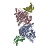
|
|---|---|
| 1 |
|
-Components
| #1: Protein | Mass: 133408.031 Da / Num. of mol.: 2 / Mutation: K-131A, N -197A, E -198A, K-287A, D-288A Source method: isolated from a genetically manipulated source Source: (gene. exp.) Human gammaherpesvirus 4 (Epstein-Barr virus)Strain: K12 / Gene: malE, b4034, JW3994, RIR1, BORF2 / Production host: Homo sapiens (human)References: UniProt: P0AEX9, UniProt: P03190, ribonucleoside-diphosphate reductase #2: Protein | Mass: 25342.541 Da / Num. of mol.: 2 Source method: isolated from a genetically manipulated source Source: (gene. exp.) Homo sapiens (human) / Gene: APOBEC3B / Production host: Homo sapiens (human)References: UniProt: Q9UH17, single-stranded DNA cytosine deaminase #3: Chemical |   Mass: 65.409 Da / Num. of mol.: 2 / Source method: obtained synthetically / Formula: Zn Mass: 65.409 Da / Num. of mol.: 2 / Source method: obtained synthetically / Formula: ZnHas ligand of interest | N | Has protein modification | Y | |
|---|
-Experimental details
-Experiment
| Experiment | Method: ELECTRON MICROSCOPY |
|---|---|
| EM experiment | Aggregation state: PARTICLE / 3D reconstruction method: single particle reconstruction |
- Sample preparation
| Component |
| ||||||||||||||||||||||||
|---|---|---|---|---|---|---|---|---|---|---|---|---|---|---|---|---|---|---|---|---|---|---|---|---|---|
| Source (natural) |
| ||||||||||||||||||||||||
| Source (recombinant) |
| ||||||||||||||||||||||||
| Buffer solution | pH: 8 | ||||||||||||||||||||||||
| Specimen | Embedding applied: NO / Shadowing applied: NO / Staining applied: NO / Vitrification applied: YES | ||||||||||||||||||||||||
| Vitrification | Cryogen name: ETHANE |
- Electron microscopy imaging
| Experimental equipment |  Model: Titan Krios / Image courtesy: FEI Company |
|---|---|
| Microscopy | Model: FEI TITAN KRIOS |
| Electron gun | Electron source:  FIELD EMISSION GUN / Accelerating voltage: 300 kV / Illumination mode: FLOOD BEAM FIELD EMISSION GUN / Accelerating voltage: 300 kV / Illumination mode: FLOOD BEAM |
| Electron lens | Mode: BRIGHT FIELD / Nominal defocus max: 2500 nm / Nominal defocus min: 800 nm |
| Image recording | Electron dose: 1.2 e/Å2 / Film or detector model: GATAN K3 BIOQUANTUM (6k x 4k) |
- Processing
| Software | Name: PHENIX / Version: 1.20.1_4486: / Classification: refinement | ||||||||||||||||||||||||
|---|---|---|---|---|---|---|---|---|---|---|---|---|---|---|---|---|---|---|---|---|---|---|---|---|---|
| EM software | Name: PHENIX / Category: model refinement | ||||||||||||||||||||||||
| CTF correction | Type: PHASE FLIPPING AND AMPLITUDE CORRECTION | ||||||||||||||||||||||||
| 3D reconstruction | Resolution: 2.55 Å / Resolution method: FSC 0.143 CUT-OFF / Num. of particles: 243192 / Symmetry type: POINT | ||||||||||||||||||||||||
| Refine LS restraints |
|
