 Movie
Movie Controller


+ Open data

- Basic information
| Entry | Database: PDB / ID: 7rrw | ||||||
|---|---|---|---|---|---|---|---|
| Title | Monomeric CRM197 expressed in E. coli | ||||||
 Components Components | Diphtheria toxin | ||||||
 Keywords Keywords | TOXIN / inactivated diphtheria toxin / vaccine carrier / E. coli expression | ||||||
| Function / homology |  Function and homology information Function and homology informationNAD+-diphthamide ADP-ribosyltransferase activity / toxin activity / extracellular space Similarity search - Function | ||||||
| Biological species |  Corynebacterium diphtheriae (bacteria) Corynebacterium diphtheriae (bacteria) | ||||||
| Method |  X-RAY DIFFRACTION / SYNCHROTRON / MOLECULAR REPLACEMENT / Resolution: 2 Å X-RAY DIFFRACTION / SYNCHROTRON / MOLECULAR REPLACEMENT / Resolution: 2 Å | ||||||
 Authors Authors | Gallagher, D.T. / Lees, A. | ||||||
| Funding support |  United States, 1items United States, 1items
| ||||||
 Citation Citation | Journal: Acta Crystallogr.,Sect.F / Year: 2023 Title: Monomeric crystal structure of the vaccine carrier protein CRM 197 and implications for vaccine development. Authors: Gallagher, D.T. / Oganesyan, N. / Lees, A. | ||||||
| History |
|
- Structure visualization
| Structure viewer | Molecule: MolmilJmol/JSmol |
|---|
- Downloads & links
-Download
| PDBx/mmCIF format | 7rrw.cif.gz | 215.1 KB | Display | PDBx/mmCIF format |
|---|---|---|---|---|
| PDB format | pdb7rrw.ent.gz | 170.1 KB | Display | PDB format |
| PDBx/mmJSON format | 7rrw.json.gz | Tree view | PDBx/mmJSON format | |
| Others |  Other downloads Other downloads |
-Validation report
| Summary document | 7rrw_validation.pdf.gz | 440 KB | Display | wwPDB validaton report |
|---|---|---|---|---|
| Full document | 7rrw_full_validation.pdf.gz | 443.2 KB | Display | |
| Data in XML | 7rrw_validation.xml.gz | 23.9 KB | Display | |
| Data in CIF | 7rrw_validation.cif.gz | 36 KB | Display | |
| Arichive directory | https://data.pdbj.org/pub/pdb/validation_reports/rr/7rrwftp://data.pdbj.org/pub/pdb/validation_reports/rr/7rrw | HTTPS FTP |
-Related structure data
| Related structure data | 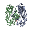 5i82S S: Starting model for refinement |
|---|---|
| Similar structure data |
-Links
 PDBj
PDBj
- Assembly
| Deposited unit | 
| ||||||||
|---|---|---|---|---|---|---|---|---|---|
| 1 |
| ||||||||
| Unit cell |
|
-Components
| #1: Protein | Mass: 58483.422 Da / Num. of mol.: 1 / Fragment: engineered variant / Mutation: G52E Source method: isolated from a genetically manipulated source Source: (gene. exp.) Corynebacterium diphtheriae (bacteria) / Plasmid: pET-15b / Production host: |
|---|---|
| #2: Chemical | ChemComp-TRS /   Mass: 122.143 Da / Num. of mol.: 1 / Source method: obtained synthetically / Formula: C4H12NO3 / Comment: pH buffer*YM Mass: 122.143 Da / Num. of mol.: 1 / Source method: obtained synthetically / Formula: C4H12NO3 / Comment: pH buffer*YM |
| #3: Water | ChemComp-HOH /  Mass: 18.015 Da / Num. of mol.: 424 / Source method: isolated from a natural source / Formula: H2O Mass: 18.015 Da / Num. of mol.: 424 / Source method: isolated from a natural source / Formula: H2O |
| Has ligand of interest | N |
| Has protein modification | Y |
-Experimental details
-Experiment
| Experiment | Method: X-RAY DIFFRACTION / Number of used crystals: 1 |
|---|
- Sample preparation
| Crystal | Density Matthews: 2.76 Å3/Da / Density % sol: 55.49 % |
|---|---|
| Crystal grow | Temperature: 295 K / Method: vapor diffusion / pH: 8 / Details: 12% (w/v) PEG 10000, 40 mM Ca Acetate |
-Data collection
| Diffraction | Mean temperature: 100 K / Serial crystal experiment: N |
|---|---|
| Diffraction source | Source: SYNCHROTRON / Site: APS / Beamline: 23-ID-D / Wavelength: 1.03317 Å |
| Detector | Type: DECTRIS PILATUS3 6M / Detector: PIXEL / Date: Jul 23, 2020 |
| Radiation | Protocol: SINGLE WAVELENGTH / Monochromatic (M) / Laue (L): M / Scattering type: x-ray |
| Radiation wavelength | Wavelength: 1.03317 Å / Relative weight: 1 |
| Reflection | Resolution: 1.7→45.71 Å / Num. obs: 87511 / % possible obs: 93.1 % / Redundancy: 4.1 % / CC1/2: 0.996 / Rmerge(I) obs: 0.082 / Rpim(I) all: 0.042 / Rrim(I) all: 0.092 / Net I/σ(I): 7.4 |
| Reflection shell | Resolution: 1.7→1.77 Å / Redundancy: 1.3 % / Rmerge(I) obs: 0.41 / Num. unique obs: 410 / Rpim(I) all: 0.913 / Rrim(I) all: 0.98 / % possible all: 77.1 |
- Processing
| Software |
| |||||||||||||||||||||||||||||||||||||||||||||
|---|---|---|---|---|---|---|---|---|---|---|---|---|---|---|---|---|---|---|---|---|---|---|---|---|---|---|---|---|---|---|---|---|---|---|---|---|---|---|---|---|---|---|---|---|---|---|
| Refinement | Method to determine structure: MOLECULAR REPLACEMENT Starting model: 5i82 Resolution: 2→16 Å / Cor.coef. Fo:Fc: 0.962 / Cor.coef. Fo:Fc free: 0.944 / SU B: 6.295 / SU ML: 0.093 / Cross valid method: THROUGHOUT / σ(F): 0 / ESU R: 0.157 / ESU R Free: 0.144 / Stereochemistry target values: MAXIMUM LIKELIHOOD / Details: U VALUES : WITH TLS ADDED
| |||||||||||||||||||||||||||||||||||||||||||||
| Solvent computation | Ion probe radii: 0.8 Å / Shrinkage radii: 0.8 Å / VDW probe radii: 1.2 Å / Solvent model: MASK | |||||||||||||||||||||||||||||||||||||||||||||
| Displacement parameters | Biso max: 111.69 Å2 / Biso mean: 35.651 Å2 / Biso min: 16.74 Å2
| |||||||||||||||||||||||||||||||||||||||||||||
| Refinement step | Cycle: final / Resolution: 2→16 Å
| |||||||||||||||||||||||||||||||||||||||||||||
| Refine LS restraints |
| |||||||||||||||||||||||||||||||||||||||||||||
| LS refinement shell | Resolution: 2→2.051 Å / Rfactor Rfree error: 0 / Total num. of bins used: 20
| |||||||||||||||||||||||||||||||||||||||||||||
| Refinement TLS params. | Method: refined / Origin x: -1.947 Å / Origin y: 32.603 Å / Origin z: 18.567 Å
| |||||||||||||||||||||||||||||||||||||||||||||
| Refinement TLS group |
|
