 Movie
Movie Controller

[English] 日本語
 Yorodumi
- PDB-7rp9: X-ray crystal structure of OXA-24/40 V130D in complex with imipenem -
+ Open data

- Basic information
| Entry | Database: PDB / ID: 7rp9 | |||||||||
|---|---|---|---|---|---|---|---|---|---|---|
| Title | X-ray crystal structure of OXA-24/40 V130D in complex with imipenem | |||||||||
 Components Components | Beta-lactamase | |||||||||
 Keywords Keywords | HYDROLASE/Inhibitor / acyl-enzyme complex / carbapenemase / HYDROLASE / HYDROLASE-Inhibitor complex | |||||||||
| Function / homology |  Function and homology information Function and homology information | |||||||||
| Biological species |  Acinetobacter baumannii (bacteria) Acinetobacter baumannii (bacteria) | |||||||||
| Method |  X-RAY DIFFRACTION / SYNCHROTRON / MOLECULAR REPLACEMENT / molecular replacement / Resolution: 1.94 Å X-RAY DIFFRACTION / SYNCHROTRON / MOLECULAR REPLACEMENT / molecular replacement / Resolution: 1.94 Å | |||||||||
 Authors Authors | Powers, R.A. / Mitchell, J.M. / June, C.M. | |||||||||
| Funding support |  United States, 2items United States, 2items
| |||||||||
 Citation Citation | Journal: J.Biol.Chem. / Year: 2022 Title: Conformational flexibility in carbapenem hydrolysis drives substrate specificity of the class D carbapenemase OXA-24/40. Authors: Mitchell, J.M. / June, C.M. / Baggett, V.L. / Lowe, B.C. / Ruble, J.F. / Bonomo, R.A. / Leonard, D.A. / Powers, R.A. | |||||||||
| History |
|
- Structure visualization
| Structure viewer | Molecule: MolmilJmol/JSmol |
|---|
- Downloads & links
-Download
| PDBx/mmCIF format | 7rp9.cif.gz | 67.8 KB | Display | PDBx/mmCIF format |
|---|---|---|---|---|
| PDB format | pdb7rp9.ent.gz | 47.4 KB | Display | PDB format |
| PDBx/mmJSON format | 7rp9.json.gz | Tree view | PDBx/mmJSON format | |
| Others |  Other downloads Other downloads |
-Validation report
| Arichive directory | https://data.pdbj.org/pub/pdb/validation_reports/rp/7rp9ftp://data.pdbj.org/pub/pdb/validation_reports/rp/7rp9 | HTTPS FTP |
|---|
-Related structure data
| Related structure data | 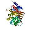 7rp8C 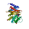 7rpaC 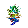 7rpbC 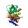 7rpcC 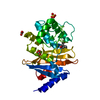 7rpdC 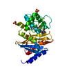 7rpeC 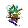 7rpfC 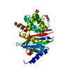 7rpgC 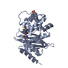 3paeS S: Starting model for refinement C: citing same article ( |
|---|---|
| Similar structure data |
-Links
 PDBj
PDBj


- Assembly
| Deposited unit | 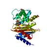
| |||||||||
|---|---|---|---|---|---|---|---|---|---|---|
| 1 |
| |||||||||
| Unit cell |
| |||||||||
| Components on special symmetry positions |
|
-Components
-Protein , 1 types, 1 molecules A
| #1: Protein | Mass: 27690.809 Da / Num. of mol.: 1 / Mutation: V130D Source method: isolated from a genetically manipulated source Source: (gene. exp.) Acinetobacter baumannii (bacteria)Gene: blaOXA-33, bla-OXA-40, blaOXA-24, blaOXA-40, oxa-24, oxa40, SI89_16690 Plasmid: pET24a / Production host: |
|---|
-Non-polymers , 5 types, 99 molecules 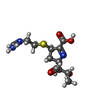
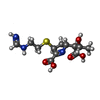


| #2: Chemical | ChemComp-ID1 /  Mass: 301.362 Da / Num. of mol.: 1 / Source method: obtained synthetically / Formula: C12H19N3O4S / Feature type: SUBJECT OF INVESTIGATION Mass: 301.362 Da / Num. of mol.: 1 / Source method: obtained synthetically / Formula: C12H19N3O4S / Feature type: SUBJECT OF INVESTIGATION |
|---|---|
| #3: Chemical | ChemComp-HIW / ( Mass: 317.361 Da / Num. of mol.: 1 / Source method: obtained synthetically / Formula: C12H19N3O5S / Feature type: SUBJECT OF INVESTIGATION Mass: 317.361 Da / Num. of mol.: 1 / Source method: obtained synthetically / Formula: C12H19N3O5S / Feature type: SUBJECT OF INVESTIGATION |
| #4: Chemical | ChemComp-SO4 /  Mass: 96.063 Da / Num. of mol.: 1 / Source method: obtained synthetically / Formula: SO4 Mass: 96.063 Da / Num. of mol.: 1 / Source method: obtained synthetically / Formula: SO4 |
| #5: Chemical | ChemComp-BCT /  Mass: 61.017 Da / Num. of mol.: 1 / Source method: isolated from a natural source / Formula: CHO3 / Comment: pH buffer*YM Mass: 61.017 Da / Num. of mol.: 1 / Source method: isolated from a natural source / Formula: CHO3 / Comment: pH buffer*YM |
| #6: Water | ChemComp-HOH / Mass: 18.015 Da / Num. of mol.: 95 / Source method: isolated from a natural source / Formula: H2O |
-Details
| Has ligand of interest | Y |
|---|---|
| Has protein modification | Y |
-Experimental details
-Experiment
| Experiment | Method: X-RAY DIFFRACTION / Number of used crystals: 1 |
|---|
- Sample preparation
| Crystal grow | Temperature: 298 K / Method: vapor diffusion, hanging drop / pH: 8.5 / Details: 0.1 M TRIS HCL, 2.0 M ammonium sulfate |
|---|
-Data collection
| Diffraction | Mean temperature: 100 K / Serial crystal experiment: N | ||||||||||||||||||||||||||||||
|---|---|---|---|---|---|---|---|---|---|---|---|---|---|---|---|---|---|---|---|---|---|---|---|---|---|---|---|---|---|---|---|
| Diffraction source | Source: SYNCHROTRON / Site: APS / Beamline: 21-ID-D / Wavelength: 1.1272 Å | ||||||||||||||||||||||||||||||
| Detector | Type: DECTRIS EIGER X 9M / Detector: PIXEL / Date: Nov 12, 2016 | ||||||||||||||||||||||||||||||
| Radiation | Protocol: SINGLE WAVELENGTH / Monochromatic (M) / Laue (L): M / Scattering type: x-ray | ||||||||||||||||||||||||||||||
| Radiation wavelength | Wavelength: 1.1272 Å / Relative weight: 1 | ||||||||||||||||||||||||||||||
| Reflection | Resolution: 1.938→66.24 Å / Num. obs: 34803 / % possible obs: 100 % / Redundancy: 7.2 % / CC1/2: 0.998 / Rmerge(I) obs: 0.103 / Rpim(I) all: 0.041 / Rrim(I) all: 0.111 / Net I/σ(I): 13.9 / Num. measured all: 249722 | ||||||||||||||||||||||||||||||
| Reflection shell | Diffraction-ID: 1
|
-Phasing
| Phasing | Method: molecular replacement |
|---|
- Processing
| Software |
| |||||||||||||||||||||||||||||||||||||||||||||
|---|---|---|---|---|---|---|---|---|---|---|---|---|---|---|---|---|---|---|---|---|---|---|---|---|---|---|---|---|---|---|---|---|---|---|---|---|---|---|---|---|---|---|---|---|---|---|
| Refinement | Method to determine structure: MOLECULAR REPLACEMENT Starting model: 3PAE Resolution: 1.94→66.24 Å / Cor.coef. Fo:Fc: 0.958 / Cor.coef. Fo:Fc free: 0.952 / SU B: 3.674 / SU ML: 0.097 / Cross valid method: THROUGHOUT / σ(F): 0 / ESU R: 0.113 / ESU R Free: 0.113 / Stereochemistry target values: MAXIMUM LIKELIHOOD Details: HYDROGENS HAVE BEEN USED IF PRESENT IN THE INPUT U VALUES : REFINED INDIVIDUALLY
| |||||||||||||||||||||||||||||||||||||||||||||
| Solvent computation | Ion probe radii: 0.8 Å / Shrinkage radii: 0.8 Å / VDW probe radii: 1.2 Å / Solvent model: BABINET MODEL WITH MASK | |||||||||||||||||||||||||||||||||||||||||||||
| Displacement parameters | Biso max: 108.39 Å2 / Biso mean: 39.278 Å2 / Biso min: 22.03 Å2
| |||||||||||||||||||||||||||||||||||||||||||||
| Refinement step | Cycle: final / Resolution: 1.94→66.24 Å
| |||||||||||||||||||||||||||||||||||||||||||||
| Refine LS restraints |
| |||||||||||||||||||||||||||||||||||||||||||||
| LS refinement shell | Resolution: 1.94→1.988 Å / Rfactor Rfree error: 0
|
