[English] 日本語
 Yorodumi
- PDB-7rod: Cryo-EM reconstruction of Sulfolobus monocaudavirus SMV1, symmetry 8 -
+ Open data
- Basic information
| Entry | Database: PDB / ID: 7rod | |||||||||||||||||||||||||||||||||
|---|---|---|---|---|---|---|---|---|---|---|---|---|---|---|---|---|---|---|---|---|---|---|---|---|---|---|---|---|---|---|---|---|---|---|
| Title | Cryo-EM reconstruction of Sulfolobus monocaudavirus SMV1, symmetry 8 | |||||||||||||||||||||||||||||||||
 Components Components | major capsid protein | |||||||||||||||||||||||||||||||||
 Keywords Keywords | VIRUS / helical symmetry / archaeal virus / lemon-shaped virus / spindle-shaped virus | |||||||||||||||||||||||||||||||||
| Function / homology | membrane / Hypothetical membrane protein Function and homology information Function and homology information | |||||||||||||||||||||||||||||||||
| Biological species |   Sulfolobus monocaudavirus SMV1 Sulfolobus monocaudavirus SMV1 | |||||||||||||||||||||||||||||||||
| Method | ELECTRON MICROSCOPY / helical reconstruction / cryo EM / Resolution: 3.8 Å | |||||||||||||||||||||||||||||||||
 Authors Authors | Wang, F. / Cvirkaite-Krupovic, V. / Krupovic, M. / Egelman, E.H. | |||||||||||||||||||||||||||||||||
| Funding support |  United States, 2items United States, 2items
| |||||||||||||||||||||||||||||||||
 Citation Citation |  Journal: Cell / Year: 2022 Journal: Cell / Year: 2022Title: Spindle-shaped archaeal viruses evolved from rod-shaped ancestors to package a larger genome. Authors: Fengbin Wang / Virginija Cvirkaite-Krupovic / Matthijn Vos / Leticia C Beltran / Mark A B Kreutzberger / Jean-Marie Winter / Zhangli Su / Jun Liu / Stefan Schouten / Mart Krupovic / Edward H Egelman /    Abstract: Spindle- or lemon-shaped viruses infect archaea in diverse environments. Due to the highly pleomorphic nature of these virions, which can be found with cylindrical tails emanating from the spindle- ...Spindle- or lemon-shaped viruses infect archaea in diverse environments. Due to the highly pleomorphic nature of these virions, which can be found with cylindrical tails emanating from the spindle-shaped body, structural studies of these capsids have been challenging. We have determined the atomic structure of the capsid of Sulfolobus monocaudavirus 1, a virus that infects hosts living in nearly boiling acid. A highly hydrophobic protein, likely integrated into the host membrane before the virions assemble, forms 7 strands that slide past each other in both the tails and the spindle body. We observe the discrete steps that occur as the tail tubes expand, and these are due to highly conserved quasiequivalent interactions with neighboring subunits maintained despite significant diameter changes. Our results show how helical assemblies can vary their diameters, becoming nearly spherical to package a larger genome and suggest how all spindle-shaped viruses have evolved from archaeal rod-like viruses. | |||||||||||||||||||||||||||||||||
| History |
|
- Structure visualization
| Structure viewer | Molecule:  Molmil Molmil Jmol/JSmol Jmol/JSmol |
|---|
- Downloads & links
- Download
| PDBx/mmCIF format |  7rod.cif.gz 7rod.cif.gz | 32 KB | Display |  PDBx/mmCIF format PDBx/mmCIF format |
|---|---|---|---|---|
| PDB format |  pdb7rod.ent.gz pdb7rod.ent.gz | 20.7 KB | Display |  PDB format PDB format |
| PDBx/mmJSON format |  7rod.json.gz 7rod.json.gz | Tree view |  PDBx/mmJSON format PDBx/mmJSON format | |
| Others |  Other downloads Other downloads |
-Validation report
| Arichive directory |  https://data.pdbj.org/pub/pdb/validation_reports/ro/7rod https://data.pdbj.org/pub/pdb/validation_reports/ro/7rod ftp://data.pdbj.org/pub/pdb/validation_reports/ro/7rod ftp://data.pdbj.org/pub/pdb/validation_reports/ro/7rod | HTTPS FTP |
|---|
-Related structure data
| Related structure data |  24592MC  7ro2C  7ro3C  7ro4C  7ro5C  7ro6C  7robC  7rocC  7roeC  7rogC  7rohC  7roiC M: map data used to model this data C: citing same article ( |
|---|---|
| Similar structure data | Similarity search - Function & homology  F&H Search F&H Search |
- Links
Links
- Assembly
| Deposited unit | 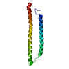
|
|---|---|
| 1 | x 203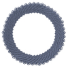
|
| 2 |
|
| 3 |
|
| Symmetry | Helical symmetry: (Circular symmetry: 7 / Dyad axis: no / N subunits divisor: 1 / Num. of operations: 203 / Rise per n subunits: 4.378 Å / Rotation per n subunits: -7.716 °) |
- Components
| #1: Protein | Mass: 16179.297 Da / Num. of mol.: 1 / Source method: isolated from a natural source / Source: (natural)   Sulfolobus monocaudavirus SMV1 / References: UniProt: W0UUV5 Sulfolobus monocaudavirus SMV1 / References: UniProt: W0UUV5 |
|---|---|
| Has protein modification | N |
-Experimental details
-Experiment
| Experiment | Method: ELECTRON MICROSCOPY |
|---|---|
| EM experiment | Aggregation state: FILAMENT / 3D reconstruction method: helical reconstruction |
- Sample preparation
| Component | Name: Sulfolobus monocaudavirus SMV1 / Type: VIRUS / Entity ID: all / Source: NATURAL |
|---|---|
| Source (natural) | Organism:   Sulfolobus monocaudavirus SMV1 Sulfolobus monocaudavirus SMV1 |
| Details of virus | Empty: NO / Enveloped: NO / Isolate: STRAIN / Type: VIRION |
| Buffer solution | pH: 6 |
| Specimen | Embedding applied: NO / Shadowing applied: NO / Staining applied: NO / Vitrification applied: YES |
| Vitrification | Cryogen name: ETHANE |
- Electron microscopy imaging
| Experimental equipment |  Model: Titan Krios / Image courtesy: FEI Company |
|---|---|
| Microscopy | Model: FEI TITAN KRIOS |
| Electron gun | Electron source:  FIELD EMISSION GUN / Accelerating voltage: 300 kV / Illumination mode: FLOOD BEAM FIELD EMISSION GUN / Accelerating voltage: 300 kV / Illumination mode: FLOOD BEAM |
| Electron lens | Mode: BRIGHT FIELD |
| Image recording | Electron dose: 50 e/Å2 / Film or detector model: GATAN K3 (6k x 4k) |
- Processing
| Software | Name: PHENIX / Version: 1.19.1_4122: / Classification: refinement | ||||||||||||||||||||||||
|---|---|---|---|---|---|---|---|---|---|---|---|---|---|---|---|---|---|---|---|---|---|---|---|---|---|
| EM software | Name: PHENIX / Category: model refinement | ||||||||||||||||||||||||
| CTF correction | Type: PHASE FLIPPING AND AMPLITUDE CORRECTION | ||||||||||||||||||||||||
| Helical symmerty | Angular rotation/subunit: -7.716 ° / Axial rise/subunit: 4.378 Å / Axial symmetry: C7 | ||||||||||||||||||||||||
| 3D reconstruction | Resolution: 3.8 Å / Resolution method: FSC 0.5 CUT-OFF / Num. of particles: 256976 / Symmetry type: HELICAL | ||||||||||||||||||||||||
| Refine LS restraints |
|
 Movie
Movie Controller


 PDBj
PDBj