Mass: 18.015 Da / Num. of mol.: 118 / Source method: isolated from a natural source / Formula: H2O
Has ligand of interest
N
-
Experimental details
-
Experiment
Experiment
Method: X-RAY DIFFRACTION / Number of used crystals: 1
-
Sample preparation
Crystal
Density Matthews: 1.82 Å3/Da / Density % sol: 32.44 %
Crystal grow
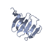
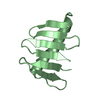
Temperature: 293 K / Method: vapor diffusion, hanging drop / pH: 8.5 Details: D.m. Plk4 PB3 was crystallized using a mother liquor (1 ml) containing 32% PEG 4000, 200 mM Li2SO4, and 200 mM Tris at pH 8.5. The hanging drop initial condition was 2 ul mother liquor plus ...Details: D.m. Plk4 PB3 was crystallized using a mother liquor (1 ml) containing 32% PEG 4000, 200 mM Li2SO4, and 200 mM Tris at pH 8.5. The hanging drop initial condition was 2 ul mother liquor plus 2 ul (15 mg/ml stock) Plk4 PB3. Crystals were transferred to a cryo condition containing mother liquor plus 20% ethylene glycol
-
Data collection
Diffraction
Mean temperature: 100 K / Serial crystal experiment: N
Type: MAR scanner 300 mm plate / Detector: IMAGE PLATE / Date: Aug 7, 2011
Radiation
Monochromator: Si (111) / Protocol: SINGLE WAVELENGTH / Monochromatic (M) / Laue (L): M / Scattering type: x-ray
Radiation wavelength
Wavelength: 1 Å / Relative weight: 1
Reflection
Resolution: 1.75→50 Å / Num. obs: 14468 / % possible obs: 93.4 % / Redundancy: 3.5 % / Biso Wilson estimate: 19.84 Å2 / Rmerge(I) obs: 0.048 / Χ2: 1.033 / Net I/σ(I): 20.2
Reflection shell
Resolution (Å)
Redundancy (%)
Rmerge(I) obs
Num. unique obs
Χ2
Diffraction-ID
% possible all
1.75-1.81
2.4
0.251
961
1.099
1
62.4
1.81-1.89
2.7
0.195
1189
1.007
1
78.8
1.89-1.97
3.3
0.141
1458
1.14
1
93.5
1.97-2.07
3.7
0.106
1531
1.077
1
99.9
2.07-2.2
3.7
0.078
1542
1.033
1
100
2.2-2.38
3.5
0.067
1538
1.045
1
99.9
2.38-2.61
3.7
0.063
1554
0.93
1
99.9
2.61-2.99
3.7
0.055
1543
0.97
1
99.9
2.99-3.77
3.7
0.041
1567
1.054
1
99.7
3.77-50
3.6
0.035
1585
1.028
1
99.6
-
Processing
Software
Name
Version
Classification
PHENIX
v1.8
refinement
HKL-2000
datascaling
PDB_EXTRACT
3.27
dataextraction
HKL-2000
datareduction
PHENIX
v1.8
phasing
Refinement
Method to determine structure: SAD / Resolution: 1.75→39.617 Å / SU ML: 0.19 / Cross valid method: THROUGHOUT / σ(F): 0 / Phase error: 25.55 / Stereochemistry target values: ML
Rfactor
Num. reflection
% reflection
Rfree
0.2327
1420
10.01 %
Rwork
0.1922
12766
-
obs
0.1964
14186
91.38 %
Solvent computation
Shrinkage radii: 0.9 Å / VDW probe radii: 1.11 Å / Solvent model: FLAT BULK SOLVENT MODEL
In the structure databanks used in Yorodumi, some data are registered as the other names, "COVID-19 virus" and "2019-nCoV". Here are the details of the virus and the list of structure data.
Jan 31, 2019. EMDB accession codes are about to change! (news from PDBe EMDB page)
EMDB accession codes are about to change! (news from PDBe EMDB page)
The allocation of 4 digits for EMDB accession codes will soon come to an end. Whilst these codes will remain in use, new EMDB accession codes will include an additional digit and will expand incrementally as the available range of codes is exhausted. The current 4-digit format prefixed with “EMD-” (i.e. EMD-XXXX) will advance to a 5-digit format (i.e. EMD-XXXXX), and so on. It is currently estimated that the 4-digit codes will be depleted around Spring 2019, at which point the 5-digit format will come into force.
The EM Navigator/Yorodumi systems omit the EMD- prefix.
Related info.:Q: What is EMD? / ID/Accession-code notation in Yorodumi/EM Navigator
Yorodumi is a browser for structure data from EMDB, PDB, SASBDB, etc.
This page is also the successor to EM Navigator detail page, and also detail information page/front-end page for Omokage search.
The word "yorodu" (or yorozu) is an old Japanese word meaning "ten thousand". "mi" (miru) is to see.
Related info.:EMDB / PDB / SASBDB / Comparison of 3 databanks / Yorodumi Search / Aug 31, 2016. New EM Navigator & Yorodumi / Yorodumi Papers / Jmol/JSmol / Function and homology information / Changes in new EM Navigator and Yorodumi
 Movie
Movie Controller


 Open data
 Basic information
 Components
 Keywords
 Function and homology information
 X-RAY DIFFRACTION /
  Authors
 United States, 1items
  Citation
 Structure visualization
 Downloads & links
 Other downloads
 PDBj
PDBj Assembly
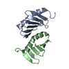

 Mass: 18.015 Da / Num. of mol.: 118 / Source method: isolated from a natural source / Formula: H2O
 Sample preparation
 Processing