 Movie
Movie Controller

[English] 日本語
 Yorodumi
- PDB-7rkb: Crystal Structure of Putative Pterin Binding Protein (PruR) from ... -
+ Open data

- Basic information
| Entry | Database: PDB / ID: 7rkb | ||||||
|---|---|---|---|---|---|---|---|
| Title | Crystal Structure of Putative Pterin Binding Protein (PruR) from Klebsiella pneumoniae in Complex with Neopterin | ||||||
 Components Components | Pterin Binding Protein | ||||||
 Keywords Keywords | PTERIN BINDING PROTEIN / Structural Genomics / Center for Structural Genomics of Infectious Diseases / CSGID / PruR | ||||||
| Function / homology | Sulfite Oxidase; Chain A, domain 2 / Oxidoreductase, molybdopterin-binding domain / Oxidoreductase, molybdopterin-binding domain / Oxidoreductase molybdopterin binding domain / Oxidoreductase, molybdopterin-binding domain superfamily / Alpha-Beta Complex / Alpha Beta / L-NEOPTERIN / Molybdopterin-dependent oxidoreductase Function and homology information Function and homology information | ||||||
| Biological species |  Klebsiella pneumoniae subsp. pneumoniae SA1 (bacteria) Klebsiella pneumoniae subsp. pneumoniae SA1 (bacteria) | ||||||
| Method |  X-RAY DIFFRACTION / SYNCHROTRON / SAD / Resolution: 2.5 Å X-RAY DIFFRACTION / SYNCHROTRON / SAD / Resolution: 2.5 Å | ||||||
 Authors Authors | Minasov, G. / Shuvalova, L. / Kiryukhina, O. / Dubrovska, I. / Satchell, K.J.F. / Center for Structural Genomics of Infectious Diseases (CSGID) | ||||||
| Funding support |  United States, 1items United States, 1items
| ||||||
 Citation Citation | Journal: Proc.Natl.Acad.Sci.USA / Year: 2024 Title: Control of biofilm formation by an Agrobacterium tumefaciens pterin-binding periplasmic protein conserved among diverse Proteobacteria. Authors: Greenwich, J.L. / Eagan, J.L. / Feirer, N. / Boswinkle, K. / Minasov, G. / Shuvalova, L. / Inniss, N.L. / Raghavaiah, J. / Ghosh, A.K. / Satchell, K.J.F. / Allen, K.D. / Fuqua, C. | ||||||
| History |
|
- Structure visualization
| Structure viewer | Molecule: MolmilJmol/JSmol |
|---|
- Downloads & links
-Download
| PDBx/mmCIF format | 7rkb.cif.gz | 77.2 KB | Display | PDBx/mmCIF format |
|---|---|---|---|---|
| PDB format | pdb7rkb.ent.gz | 57.2 KB | Display | PDB format |
| PDBx/mmJSON format | 7rkb.json.gz | Tree view | PDBx/mmJSON format | |
| Others |  Other downloads Other downloads |
-Validation report
| Arichive directory | https://data.pdbj.org/pub/pdb/validation_reports/rk/7rkbftp://data.pdbj.org/pub/pdb/validation_reports/rk/7rkb | HTTPS FTP |
|---|
-Related structure data
| Related structure data | 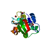 7komC 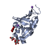 7kosC 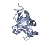 7kouC 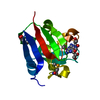 7kp2C C: citing same article ( |
|---|---|
| Similar structure data | |
| Other databases |
-Links
 PDBj
PDBj- Assembly
| Deposited unit | 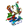
| ||||||||
|---|---|---|---|---|---|---|---|---|---|
| 1 |
| ||||||||
| Unit cell |
|
-Components
| #1: Protein | Mass: 17068.654 Da / Num. of mol.: 1 Source method: isolated from a genetically manipulated source Source: (gene. exp.) Klebsiella pneumoniae subsp. pneumoniae SA1 (bacteria)Gene: RJA_09115 / Plasmid: pMCSG53 / Production host: | ||||||||
|---|---|---|---|---|---|---|---|---|---|
| #2: Chemical | ChemComp-NEU / 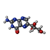  Mass: 253.215 Da / Num. of mol.: 1 / Source method: obtained synthetically / Formula: C9H11N5O4 / Feature type: SUBJECT OF INVESTIGATION Mass: 253.215 Da / Num. of mol.: 1 / Source method: obtained synthetically / Formula: C9H11N5O4 / Feature type: SUBJECT OF INVESTIGATION | ||||||||
| #3: Chemical | ChemComp-CL /   Mass: 35.453 Da / Num. of mol.: 5 / Source method: obtained synthetically / Formula: Cl Mass: 35.453 Da / Num. of mol.: 5 / Source method: obtained synthetically / Formula: Cl#4: Chemical | ChemComp-SO4 /   Mass: 96.063 Da / Num. of mol.: 4 / Source method: obtained synthetically / Formula: SO4 Mass: 96.063 Da / Num. of mol.: 4 / Source method: obtained synthetically / Formula: SO4#5: Water | ChemComp-HOH / |  Mass: 18.015 Da / Num. of mol.: 20 / Source method: isolated from a natural source / Formula: H2O Mass: 18.015 Da / Num. of mol.: 20 / Source method: isolated from a natural source / Formula: H2OHas ligand of interest | Y | Has protein modification | Y | |
-Experimental details
-Experiment
| Experiment | Method: X-RAY DIFFRACTION / Number of used crystals: 1 |
|---|
- Sample preparation
| Crystal | Density Matthews: 2.34 Å3/Da / Density % sol: 47.53 % |
|---|---|
| Crystal grow | Temperature: 292 K / Method: vapor diffusion, sitting drop / pH: 8.5 Details: 8.0 mg/mL protein in 0.5 M sodium chloride, 0.01 M Tris, pH 8.3, 2 mM Neopterin against Classics II screen G5 (0.2 M lithium sulfate, 0.1 M Tris, pH 8.5, 25% w/v PEG3350) |
-Data collection
| Diffraction | Mean temperature: 100 K / Serial crystal experiment: N |
|---|---|
| Diffraction source | Source: SYNCHROTRON / Site: APS / Beamline: 21-ID-E / Wavelength: 0.97872 Å |
| Detector | Type: MARMOSAIC 300 mm CCD / Detector: CCD / Date: Jun 5, 2019 / Details: Be |
| Radiation | Monochromator: C(111) / Protocol: SINGLE WAVELENGTH / Monochromatic (M) / Laue (L): M / Scattering type: x-ray |
| Radiation wavelength | Wavelength: 0.97872 Å / Relative weight: 1 |
| Reflection | Resolution: 2.5→30 Å / Num. obs: 5990 / % possible obs: 100 % / Observed criterion σ(I): -3 / Redundancy: 13.7 % / Biso Wilson estimate: 57 Å2 / CC1/2: 1 / CC star: 1 / Rmerge(I) obs: 0.157 / Rpim(I) all: 0.045 / Rrim(I) all: 0.163 / Rsym value: 0.157 / Χ2: 1.261 / Net I/σ(I): 20.1 |
| Reflection shell | Resolution: 2.5→2.54 Å / Redundancy: 14.2 % / Rmerge(I) obs: 1.675 / Mean I/σ(I) obs: 2.2 / Num. unique obs: 298 / CC1/2: 0.709 / CC star: 0.911 / Rpim(I) all: 0.461 / Rsym value: 1.675 / Χ2: 1.004 / % possible all: 100 |
- Processing
| Software |
| |||||||||||||||||||||||||||||||||||||||||||||||||||||||||||||||||||||||||||||||||||||||||||||||||||||||||||||||||||||||||||||
|---|---|---|---|---|---|---|---|---|---|---|---|---|---|---|---|---|---|---|---|---|---|---|---|---|---|---|---|---|---|---|---|---|---|---|---|---|---|---|---|---|---|---|---|---|---|---|---|---|---|---|---|---|---|---|---|---|---|---|---|---|---|---|---|---|---|---|---|---|---|---|---|---|---|---|---|---|---|---|---|---|---|---|---|---|---|---|---|---|---|---|---|---|---|---|---|---|---|---|---|---|---|---|---|---|---|---|---|---|---|---|---|---|---|---|---|---|---|---|---|---|---|---|---|---|---|---|
| Refinement | Method to determine structure: SAD / Resolution: 2.5→29.53 Å / Cor.coef. Fo:Fc: 0.959 / Cor.coef. Fo:Fc free: 0.953 / SU B: 21.645 / SU ML: 0.227 / Cross valid method: THROUGHOUT / σ(F): 0 / ESU R: 0.827 / ESU R Free: 0.29 / Stereochemistry target values: MAXIMUM LIKELIHOOD Details: HYDROGENS HAVE BEEN ADDED IN THE RIDING POSITIONS U VALUES : WITH TLS ADDED
| |||||||||||||||||||||||||||||||||||||||||||||||||||||||||||||||||||||||||||||||||||||||||||||||||||||||||||||||||||||||||||||
| Solvent computation | Ion probe radii: 0.8 Å / Shrinkage radii: 0.8 Å / VDW probe radii: 1.2 Å / Solvent model: MASK | |||||||||||||||||||||||||||||||||||||||||||||||||||||||||||||||||||||||||||||||||||||||||||||||||||||||||||||||||||||||||||||
| Displacement parameters | Biso max: 175.73 Å2 / Biso mean: 68.845 Å2 / Biso min: 39.13 Å2
| |||||||||||||||||||||||||||||||||||||||||||||||||||||||||||||||||||||||||||||||||||||||||||||||||||||||||||||||||||||||||||||
| Refinement step | Cycle: final / Resolution: 2.5→29.53 Å
| |||||||||||||||||||||||||||||||||||||||||||||||||||||||||||||||||||||||||||||||||||||||||||||||||||||||||||||||||||||||||||||
| Refine LS restraints |
| |||||||||||||||||||||||||||||||||||||||||||||||||||||||||||||||||||||||||||||||||||||||||||||||||||||||||||||||||||||||||||||
| LS refinement shell | Resolution: 2.502→2.566 Å / Rfactor Rfree error: 0 / Total num. of bins used: 20
| |||||||||||||||||||||||||||||||||||||||||||||||||||||||||||||||||||||||||||||||||||||||||||||||||||||||||||||||||||||||||||||
| Refinement TLS params. | Method: refined / Refine-ID: X-RAY DIFFRACTION
| |||||||||||||||||||||||||||||||||||||||||||||||||||||||||||||||||||||||||||||||||||||||||||||||||||||||||||||||||||||||||||||
| Refinement TLS group |
|
