- PDB-7rk3: Crystal structure of human N-myristoyltransferase 1 fragment (res... -
+
Open data
ID or keywords:
Loading...
-
Basic information
Entry
Database: PDB / ID: 7rk3
Title
Crystal structure of human N-myristoyltransferase 1 fragment (residues 109-496) bound to diacylated human Arf6 octapeptide and Coenzyme A
Components
ADP-ribosylation factor 6
Glycylpeptide N-tetradecanoyltransferase 1
Keywords
TRANSFERASE
Function / homology
Function and homology information
erythrocyte apoptotic process / maintenance of postsynaptic density structure / protein localization to cleavage furrow / myristoyltransferase activity / N-terminal peptidyl-glycine N-myristoylation / peptidyl-lysine N6-myristoyltransferase activity / Late Phase of HIV Life Cycle / positive regulation of mitotic cytokinetic process / ketone metabolic process / establishment of epithelial cell polarity ...erythrocyte apoptotic process / maintenance of postsynaptic density structure / protein localization to cleavage furrow / myristoyltransferase activity / N-terminal peptidyl-glycine N-myristoylation / peptidyl-lysine N6-myristoyltransferase activity / Late Phase of HIV Life Cycle / positive regulation of mitotic cytokinetic process / ketone metabolic process / establishment of epithelial cell polarity / regulation of opsin-mediated signaling pathway / regulation of dendritic spine development / Activation, myristolyation of BID and translocation to mitochondria / positive regulation of protein localization to mitochondrion / negative regulation of protein localization to cell surface / protein localization to endosome / regulation of Rac protein signal transduction / negative regulation of dendrite development / ruffle assembly / glycylpeptide N-tetradecanoyltransferase / glycylpeptide N-tetradecanoyltransferase activity / positive regulation of keratinocyte migration / regulation of filopodium assembly / positive regulation of focal adhesion disassembly / endocytic recycling / MET receptor recycling / thioesterase binding / Flemming body / filopodium membrane / TBC/RABGAPs / protein localization to cell surface / cortical actin cytoskeleton organization / protein localization to membrane / hepatocyte apoptotic process / positive regulation of actin filament polymerization / cleavage furrow / endocytic vesicle / regulation of presynapse assembly / synaptic vesicle endocytosis / ruffle / eNOS activation / vesicle-mediated transport / Transferases; Acyltransferases; Transferring groups other than aminoacyl groups / signaling adaptor activity / small monomeric GTPase / protein localization to plasma membrane / positive regulation of protein localization to plasma membrane / positive regulation of protein secretion / liver development / negative regulation of receptor-mediated endocytosis / cellular response to nerve growth factor stimulus / positive regulation of neuron projection development / intracellular protein transport / recycling endosome membrane / GDP binding / nervous system development / Inactivation, recovery and regulation of the phototransduction cascade / Clathrin-mediated endocytosis / presynapse / G protein activity / early endosome membrane / midbody / cell cortex / in utero embryonic development / cell differentiation / cell adhesion / endosome / postsynapse / cell division / focal adhesion / GTPase activity / GTP binding / glutamatergic synapse / Golgi apparatus / extracellular exosome / membrane / plasma membrane / cytoplasm / cytosol Similarity search - Function
ADP-ribosylation factor 6 / Glycylpeptide N-tetradecanoyltransferase, conserved site / Myristoyl-CoA:protein N-myristoyltransferase signature 1. / Myristoyl-CoA:protein N-myristoyltransferase signature 2. / Glycylpeptide N-tetradecanoyltransferase / Glycylpeptide N-tetradecanoyltransferase, N-terminal / Glycylpeptide N-tetradecanoyltransferase, C-terminal / Myristoyl-CoA:protein N-myristoyltransferase, N-terminal domain / Myristoyl-CoA:protein N-myristoyltransferase, C-terminal domain / Small GTPase superfamily, ARF type ...ADP-ribosylation factor 6 / Glycylpeptide N-tetradecanoyltransferase, conserved site / Myristoyl-CoA:protein N-myristoyltransferase signature 1. / Myristoyl-CoA:protein N-myristoyltransferase signature 2. / Glycylpeptide N-tetradecanoyltransferase / Glycylpeptide N-tetradecanoyltransferase, N-terminal / Glycylpeptide N-tetradecanoyltransferase, C-terminal / Myristoyl-CoA:protein N-myristoyltransferase, N-terminal domain / Myristoyl-CoA:protein N-myristoyltransferase, C-terminal domain / Small GTPase superfamily, ARF type / Small GTPase Arf domain profile. / Sar1p-like members of the Ras-family of small GTPases / Small GTPase superfamily, ARF/SAR type / ADP-ribosylation factor family / ARF-like small GTPases; ARF, ADP-ribosylation factor / Acyl-CoA N-acyltransferase / Rab subfamily of small GTPases / Small GTP-binding protein domain / P-loop containing nucleoside triphosphate hydrolase Similarity search - Domain/homology
National Institutes of Health/National Institute of Diabetes and Digestive and Kidney Disease (NIH/NIDDK)
DK107868-04
United States
National Institutes of Health/National Institute of General Medical Sciences (NIH/NIGMS)
GM124165-01
United States
National Institutes of Health/Office of the Director
S10OD021527
Department of Energy (DOE, United States)
DE-AC02-06CH11357
United States
Citation
Journal: To Be Published Title: Crystal structure of human N-myristoyltransferase 1 fragment (residues 109-496) bound to diacylated human Arf6 octapeptide and Coenzyme A Authors: Fenwick, M.K. / Lin, H.
In the structure databanks used in Yorodumi, some data are registered as the other names, "COVID-19 virus" and "2019-nCoV". Here are the details of the virus and the list of structure data.
Jan 31, 2019. EMDB accession codes are about to change! (news from PDBe EMDB page)
EMDB accession codes are about to change! (news from PDBe EMDB page)
The allocation of 4 digits for EMDB accession codes will soon come to an end. Whilst these codes will remain in use, new EMDB accession codes will include an additional digit and will expand incrementally as the available range of codes is exhausted. The current 4-digit format prefixed with “EMD-” (i.e. EMD-XXXX) will advance to a 5-digit format (i.e. EMD-XXXXX), and so on. It is currently estimated that the 4-digit codes will be depleted around Spring 2019, at which point the 5-digit format will come into force.
The EM Navigator/Yorodumi systems omit the EMD- prefix.
Related info.:Q: What is EMD? / ID/Accession-code notation in Yorodumi/EM Navigator
Yorodumi is a browser for structure data from EMDB, PDB, SASBDB, etc.
This page is also the successor to EM Navigator detail page, and also detail information page/front-end page for Omokage search.
The word "yorodu" (or yorozu) is an old Japanese word meaning "ten thousand". "mi" (miru) is to see.
Related info.:EMDB / PDB / SASBDB / Comparison of 3 databanks / Yorodumi Search / Aug 31, 2016. New EM Navigator & Yorodumi / Yorodumi Papers / Jmol/JSmol / Function and homology information / Changes in new EM Navigator and Yorodumi
 Movie
Movie Controller

 Yorodumi
 Open data
 Basic information
 Components
 Keywords
 Function and homology information
 Homo sapiens (human)
 X-RAY DIFFRACTION /
  Authors
 United States, 4items
  Citation
 Structure visualization
 Downloads & links
 Other downloads
 PDBj
PDBj


 Assembly
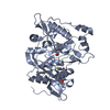


 Mass: 438.328 Da / Num. of mol.: 1 / Source method: obtained synthetically / Formula: C11H24N2O10P2S / Feature type: SUBJECT OF INVESTIGATION
 Mass: 228.371 Da / Num. of mol.: 1 / Source method: obtained synthetically / Formula: C14H28O2 / Feature type: SUBJECT OF INVESTIGATION
 Mass: 116.158 Da / Num. of mol.: 1 / Source method: obtained synthetically / Formula: C6H12O2 / Feature type: SUBJECT OF INVESTIGATION
 Sample preparation
 Processing