 Movie
Movie Controller


+ Open data

- Basic information
| Entry | Database: PDB / ID: 7rib | ||||||
|---|---|---|---|---|---|---|---|
| Title | Griffithsin mutant Y28F/Y68F/Y110F | ||||||
 Components Components | Griffithsin | ||||||
 Keywords Keywords | SUGAR BINDING PROTEIN / complex | ||||||
| Function / homology |  Function and homology information Function and homology informationN-acetylgalactosamine binding / D-glucose binding / D-mannose binding / carbohydrate binding / identical protein binding Similarity search - Function | ||||||
| Biological species |  Griffithsia sp. (eukaryote) Griffithsia sp. (eukaryote) | ||||||
| Method |  X-RAY DIFFRACTION / MOLECULAR REPLACEMENT / Resolution: 2.1 Å X-RAY DIFFRACTION / MOLECULAR REPLACEMENT / Resolution: 2.1 Å | ||||||
 Authors Authors | Zhao, G. / Sun, J. / Bewley, C.A. | ||||||
 Citation Citation | Journal: Acs Chem.Biol. / Year: 2022 Title: C 3 -Symmetric Aromatic Core of Griffithsin Is Essential for Potent Anti-HIV Activity. Authors: Sun, J. / Zhao, G. / Bylund, T. / Lee, M. / Adibhatla, S. / Kwong, P.D. / Chuang, G.Y. / Rawi, R. / Bewley, C.A. | ||||||
| History |
|
- Structure visualization
| Structure viewer | Molecule: MolmilJmol/JSmol |
|---|
- Downloads & links
-Download
| PDBx/mmCIF format | 7rib.cif.gz | 114.7 KB | Display | PDBx/mmCIF format |
|---|---|---|---|---|
| PDB format | pdb7rib.ent.gz | 70.2 KB | Display | PDB format |
| PDBx/mmJSON format | 7rib.json.gz | Tree view | PDBx/mmJSON format | |
| Others |  Other downloads Other downloads |
-Validation report
| Arichive directory | https://data.pdbj.org/pub/pdb/validation_reports/ri/7ribftp://data.pdbj.org/pub/pdb/validation_reports/ri/7rib | HTTPS FTP |
|---|
-Related structure data
| Related structure data | 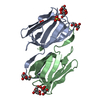 7riaC 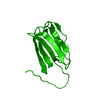 7ricC 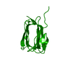 7ridC 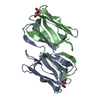 7rkgC 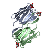 7rkiC  2gucS S: Starting model for refinement C: citing same article ( |
|---|---|
| Similar structure data |
-Links
 PDBj
PDBj


- Assembly
| Deposited unit | 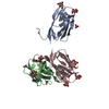
| ||||||||||||
|---|---|---|---|---|---|---|---|---|---|---|---|---|---|
| 1 | 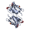
| ||||||||||||
| 2 | 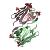
| ||||||||||||
| Unit cell |
| ||||||||||||
| Components on special symmetry positions |
|
-Components
| #1: Protein | Mass: 14770.099 Da / Num. of mol.: 3 / Mutation: Y28F, Y68F, Y110F Source method: isolated from a genetically manipulated source Source: (gene. exp.) Griffithsia sp. (strain Q66D336) (eukaryote)Strain: Q66D336 / Production host:  #2: Sugar | ChemComp-MAN /   Type: D-saccharide, alpha linking / Mass: 180.156 Da / Num. of mol.: 6 / Source method: obtained synthetically / Formula: C6H12O6 / Feature type: SUBJECT OF INVESTIGATION Type: D-saccharide, alpha linking / Mass: 180.156 Da / Num. of mol.: 6 / Source method: obtained synthetically / Formula: C6H12O6 / Feature type: SUBJECT OF INVESTIGATION#3: Chemical |   Mass: 96.063 Da / Num. of mol.: 3 / Source method: obtained synthetically / Formula: SO4 Mass: 96.063 Da / Num. of mol.: 3 / Source method: obtained synthetically / Formula: SO4#4: Water | ChemComp-HOH / |  Mass: 18.015 Da / Num. of mol.: 465 / Source method: isolated from a natural source / Formula: H2O Mass: 18.015 Da / Num. of mol.: 465 / Source method: isolated from a natural source / Formula: H2OHas ligand of interest | Y | |
|---|
-Experimental details
-Experiment
| Experiment | Method: X-RAY DIFFRACTION / Number of used crystals: 1 |
|---|
- Sample preparation
| Crystal | Density Matthews: 2.01 Å3/Da / Density % sol: 38.73 % |
|---|---|
| Crystal grow | Temperature: 291 K / Method: vapor diffusion, hanging drop Details: 0.1M bis-tris buffer, pH 6.5, 1.0 M ammonium sulfate |
-Data collection
| Diffraction | Mean temperature: 95 K / Serial crystal experiment: N |
|---|---|
| Diffraction source | Source: ROTATING ANODE / Type: RIGAKU R-AXIS IV / Wavelength: 1.54 Å |
| Detector | Type: RIGAKU SATURN A200 / Detector: CCD / Date: May 1, 2018 |
| Radiation | Protocol: SINGLE WAVELENGTH / Monochromatic (M) / Laue (L): M / Scattering type: x-ray |
| Radiation wavelength | Wavelength: 1.54 Å / Relative weight: 1 |
| Reflection | Resolution: 2.1→25 Å / Num. obs: 20011 / % possible obs: 96.4 % / Redundancy: 3.7 % / Biso Wilson estimate: 16.41 Å2 / Rmerge(I) obs: 0.2 / Net I/σ(I): 4.8 |
| Reflection shell | Resolution: 2.1→2.15 Å / Redundancy: 3.8 % / Rmerge(I) obs: 0.095 / Mean I/σ(I) obs: 1.3 / Num. unique obs: 964 / % possible all: 94.4 |
- Processing
| Software |
| |||||||||||||||||||||||||||||||||||||||||||||||||||||||||||||||||||||||||||||||||||||||||||||||||||||||||
|---|---|---|---|---|---|---|---|---|---|---|---|---|---|---|---|---|---|---|---|---|---|---|---|---|---|---|---|---|---|---|---|---|---|---|---|---|---|---|---|---|---|---|---|---|---|---|---|---|---|---|---|---|---|---|---|---|---|---|---|---|---|---|---|---|---|---|---|---|---|---|---|---|---|---|---|---|---|---|---|---|---|---|---|---|---|---|---|---|---|---|---|---|---|---|---|---|---|---|---|---|---|---|---|---|---|---|
| Refinement | Method to determine structure: MOLECULAR REPLACEMENT Starting model: 2GUC Resolution: 2.1→24.98 Å / SU ML: 0.1907 / Cross valid method: FREE R-VALUE / σ(F): 1.36 / Phase error: 20.428 Stereochemistry target values: GeoStd + Monomer Library + CDL v1.2
| |||||||||||||||||||||||||||||||||||||||||||||||||||||||||||||||||||||||||||||||||||||||||||||||||||||||||
| Solvent computation | Shrinkage radii: 0.9 Å / VDW probe radii: 1.11 Å / Solvent model: FLAT BULK SOLVENT MODEL | |||||||||||||||||||||||||||||||||||||||||||||||||||||||||||||||||||||||||||||||||||||||||||||||||||||||||
| Displacement parameters | Biso mean: 16.73 Å2 | |||||||||||||||||||||||||||||||||||||||||||||||||||||||||||||||||||||||||||||||||||||||||||||||||||||||||
| Refinement step | Cycle: LAST / Resolution: 2.1→24.98 Å
| |||||||||||||||||||||||||||||||||||||||||||||||||||||||||||||||||||||||||||||||||||||||||||||||||||||||||
| Refine LS restraints |
| |||||||||||||||||||||||||||||||||||||||||||||||||||||||||||||||||||||||||||||||||||||||||||||||||||||||||
| LS refinement shell |
|
