| 登録情報 | データベース: PDB / ID: 7rh1
|
|---|
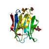
| タイトル | Crystal structure of human galectin-3 CRD in complex with Methyl 2-O-(3-nitro-benzoyl)-3-toluoyl-b-D-talopyranoside |
|---|
 要素 要素 | Galectin-3 |
|---|
 キーワード キーワード | SUGAR BINDING PROTEIN / Carbohydrates-binding protein |
|---|
| 機能・相同性 |  機能・相同性情報 機能・相同性情報
negative regulation of NK T cell activation / negative regulation of immunological synapse formation / disaccharide binding / negative regulation of T cell activation via T cell receptor contact with antigen bound to MHC molecule on antigen presenting cell / RUNX2 regulates genes involved in differentiation of myeloid cells / regulation of T cell apoptotic process / mononuclear cell migration / receptor ligand inhibitor activity / positive regulation of mononuclear cell migration / negative regulation of endocytosis ...negative regulation of NK T cell activation / negative regulation of immunological synapse formation / disaccharide binding / negative regulation of T cell activation via T cell receptor contact with antigen bound to MHC molecule on antigen presenting cell / RUNX2 regulates genes involved in differentiation of myeloid cells / regulation of T cell apoptotic process / mononuclear cell migration / receptor ligand inhibitor activity / positive regulation of mononuclear cell migration / negative regulation of endocytosis / IgE binding / eosinophil chemotaxis / regulation of extrinsic apoptotic signaling pathway via death domain receptors / RUNX1 regulates transcription of genes involved in differentiation of myeloid cells / signaling receptor inhibitor activity / negative regulation of T cell receptor signaling pathway / protein phosphatase inhibitor activity / positive chemotaxis / positive regulation of calcium ion import / chemoattractant activity / macrophage chemotaxis / monocyte chemotaxis / regulation of T cell proliferation / Advanced glycosylation endproduct receptor signaling / ficolin-1-rich granule membrane / immunological synapse / laminin binding / neutrophil chemotaxis / epithelial cell differentiation / RNA splicing / secretory granule membrane / negative regulation of extrinsic apoptotic signaling pathway / positive regulation of protein localization to plasma membrane / spliceosomal complex / positive regulation of protein-containing complex assembly / molecular condensate scaffold activity / mRNA processing / : / carbohydrate binding / protein phosphatase binding / mitochondrial inner membrane / innate immune response / Neutrophil degranulation / cell surface / extracellular space / RNA binding / extracellular exosome / extracellular region / nucleoplasm / nucleus / membrane / plasma membrane / cytosol / cytoplasm類似検索 - 分子機能 Galectin-like / Galactoside-binding lectin / Galectin / Galectin, carbohydrate recognition domain / Galactoside-binding lectin / Galactoside-binding lectin (galectin) domain profile. / Jelly Rolls - #200 / Concanavalin A-like lectin/glucanase domain superfamily / Jelly Rolls / Sandwich / Mainly Beta類似検索 - ドメイン・相同性 |
|---|
| 生物種 |  Homo sapiens (ヒト) Homo sapiens (ヒト) |
|---|
| 手法 |  X線回折 / 分子置換 / 解像度: 1.444 Å X線回折 / 分子置換 / 解像度: 1.444 Å |
|---|
 データ登録者 データ登録者 | Collins, P.M. / Kishor, C. / Blanchard, H. |
|---|
 引用 引用 | ジャーナル: J.Med.Chem. / 年: 2022
タイトル: Novel Selective Galectin-3 Antagonists Are Cytotoxic to Acute Lymphoblastic Leukemia.
著者: Bum-Erdene, K. / Collins, P.M. / Hugo, M.W. / Tarighat, S.S. / Fei, F. / Kishor, C. / Leffler, H. / Nilsson, U.J. / Groffen, J. / Grice, I.D. / Heisterkamp, N. / Blanchard, H. |
|---|
| 履歴 | | 登録 | 2021年7月16日 | 登録サイト: RCSB / 処理サイト: RCSB |
|---|
| 改定 1.0 | 2022年7月13日 | Provider: repository / タイプ: Initial release |
|---|
| 改定 1.1 | 2023年10月18日 | Group: Data collection / Refinement description
カテゴリ: chem_comp_atom / chem_comp_bond / pdbx_initial_refinement_model |
|---|
|
|---|
 ムービー
ムービー コントローラー


 データを開く
 基本情報
基本情報 構造の表示
構造の表示 ダウンロードとリンク
 その他のダウンロード


 PDBj
PDBj


 集合体
集合体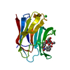


 分子量: 35.453 Da / 分子数: 2 / 由来タイプ: 合成 / 式: Cl
 分子量: 461.419 Da / 分子数: 1 / 由来タイプ: 合成 / 式: C22H23NO10 / タイプ: SUBJECT OF INVESTIGATION
 分子量: 18.015 Da / 分子数: 180 / 由来タイプ: 天然 / 式: H2O
 試料調製
試料調製 解析
解析