 Movie
Movie Controller

[English] 日本語
 Yorodumi
- PDB-7rdu: Crystal structure of Campylobacter jejuni keto said reductoisomer... -
+ Open data

- Basic information
| Entry | Database: PDB / ID: 7rdu | ||||||
|---|---|---|---|---|---|---|---|
| Title | Crystal structure of Campylobacter jejuni keto said reductoisomerase in complex with magnesium and oxidixized and reduced NADPH | ||||||
 Components Components | Ketol-acid reductoisomerase (NADP(+)) | ||||||
 Keywords Keywords | OXIDOREDUCTASE / magnesium dependent / NADPH dependent / amino acid biosynthesis / biofuel production / dodecamer | ||||||
| Function / homology |  Function and homology information Function and homology informationketol-acid reductoisomerase (NADP+) / ketol-acid reductoisomerase activity / L-valine biosynthetic process / : / NADP binding / magnesium ion binding / cytosol Similarity search - Function | ||||||
| Biological species |   Campylobacter jejuni (Campylobacter) Campylobacter jejuni (Campylobacter) | ||||||
| Method |  X-RAY DIFFRACTION / SYNCHROTRON / MOLECULAR REPLACEMENT / Resolution: 2.502 Å X-RAY DIFFRACTION / SYNCHROTRON / MOLECULAR REPLACEMENT / Resolution: 2.502 Å | ||||||
 Authors Authors | Guddat, L.W. / You, L. | ||||||
| Funding support |  Australia, 1items Australia, 1items
| ||||||
 Citation Citation | Journal: To Be Published Title: Multi-faceted approach for the engineering of enzyme variants with improved properties for industrial applications Authors: Lv, Y. / Guddat, L. | ||||||
| History |
|
- Structure visualization
| Structure viewer | Molecule: MolmilJmol/JSmol |
|---|
- Downloads & links
-Download
| PDBx/mmCIF format | 7rdu.cif.gz | 82.8 KB | Display | PDBx/mmCIF format |
|---|---|---|---|---|
| PDB format | pdb7rdu.ent.gz | 59.4 KB | Display | PDB format |
| PDBx/mmJSON format | 7rdu.json.gz | Tree view | PDBx/mmJSON format | |
| Others |  Other downloads Other downloads |
-Validation report
| Arichive directory | https://data.pdbj.org/pub/pdb/validation_reports/rd/7rduftp://data.pdbj.org/pub/pdb/validation_reports/rd/7rdu | HTTPS FTP |
|---|
-Related structure data
| Related structure data | 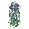 4ypoS S: Starting model for refinement |
|---|---|
| Similar structure data |
-Links
 PDBj
PDBj


- Assembly
| Deposited unit | 
| ||||||||
|---|---|---|---|---|---|---|---|---|---|
| 1 | 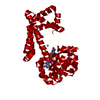
| ||||||||
| Unit cell |
| ||||||||
| Components on special symmetry positions |
|
-Components
| #1: Protein | Mass: 35737.031 Da / Num. of mol.: 1 Source method: isolated from a genetically manipulated source Source: (gene. exp.) Campylobacter jejuni (strain RM1221) (Campylobacter)Strain: RM1221 / Gene: ilvC, CJE0735 / Production host: References: UniProt: Q5HVD9, ketol-acid reductoisomerase (NADP+) | ||||||||
|---|---|---|---|---|---|---|---|---|---|
| #2: Chemical |   Mass: 24.305 Da / Num. of mol.: 2 / Source method: obtained synthetically / Formula: Mg / Feature type: SUBJECT OF INVESTIGATION Mass: 24.305 Da / Num. of mol.: 2 / Source method: obtained synthetically / Formula: Mg / Feature type: SUBJECT OF INVESTIGATION#3: Chemical | ChemComp-NAP / |   Mass: 743.405 Da / Num. of mol.: 1 / Source method: obtained synthetically / Formula: C21H28N7O17P3 / Feature type: SUBJECT OF INVESTIGATION Mass: 743.405 Da / Num. of mol.: 1 / Source method: obtained synthetically / Formula: C21H28N7O17P3 / Feature type: SUBJECT OF INVESTIGATION#4: Chemical | ChemComp-NDP / |   Mass: 745.421 Da / Num. of mol.: 1 / Source method: obtained synthetically / Formula: C21H30N7O17P3 / Feature type: SUBJECT OF INVESTIGATION Mass: 745.421 Da / Num. of mol.: 1 / Source method: obtained synthetically / Formula: C21H30N7O17P3 / Feature type: SUBJECT OF INVESTIGATION#5: Water | ChemComp-HOH / |  Mass: 18.015 Da / Num. of mol.: 37 / Source method: isolated from a natural source / Formula: H2O Mass: 18.015 Da / Num. of mol.: 37 / Source method: isolated from a natural source / Formula: H2OHas ligand of interest | Y | |
-Experimental details
-Experiment
| Experiment | Method: X-RAY DIFFRACTION / Number of used crystals: 1 |
|---|
- Sample preparation
| Crystal | Density Matthews: 2.57 Å3/Da / Density % sol: 52.1 % |
|---|---|
| Crystal grow | Temperature: 293 K / Method: vapor diffusion, hanging drop Details: The buffer was 0.2 M KNO3, 20% PEG3350. The well solution (which was not added to the drop) was 35% PEG3350, 250 mM MgCl2, 20 mM Tris-HCl pH 8.0. |
-Data collection
| Diffraction | Mean temperature: 100 K / Serial crystal experiment: N |
|---|---|
| Diffraction source | Source: SYNCHROTRON / Site: Australian Synchrotron / Beamline: MX1 / Wavelength: 1 Å |
| Detector | Type: ADSC QUANTUM 210r / Detector: CCD / Date: Jul 15, 2015 |
| Radiation | Protocol: SINGLE WAVELENGTH / Monochromatic (M) / Laue (L): M / Scattering type: x-ray |
| Radiation wavelength | Wavelength: 1 Å / Relative weight: 1 |
| Reflection | Resolution: 2.5→46.1 Å / Num. obs: 12712 / % possible obs: 99.2 % / Redundancy: 13.1 % / Rmerge(I) obs: 0.127 / Rpim(I) all: 0.039 / Net I/σ(I): 35.3 |
| Reflection shell | Resolution: 2.5→2.54 Å / Redundancy: 7 % / Rmerge(I) obs: 0.823 / Mean I/σ(I) obs: 1.4 / Num. unique obs: 554 / Rpim(I) all: 0.603 / % possible all: 91.3 |
- Processing
| Software |
| ||||||||||||||||||||||||||||||||||||||||||||||||||||||||||||||||||||||
|---|---|---|---|---|---|---|---|---|---|---|---|---|---|---|---|---|---|---|---|---|---|---|---|---|---|---|---|---|---|---|---|---|---|---|---|---|---|---|---|---|---|---|---|---|---|---|---|---|---|---|---|---|---|---|---|---|---|---|---|---|---|---|---|---|---|---|---|---|---|---|---|
| Refinement | Method to determine structure: MOLECULAR REPLACEMENT Starting model: 4YPO Resolution: 2.502→46 Å / SU ML: 0.38 / Cross valid method: FREE R-VALUE / σ(F): 1.33 / Phase error: 29.09 / Stereochemistry target values: ML
| ||||||||||||||||||||||||||||||||||||||||||||||||||||||||||||||||||||||
| Solvent computation | Shrinkage radii: 0.9 Å / VDW probe radii: 1.11 Å / Solvent model: FLAT BULK SOLVENT MODEL | ||||||||||||||||||||||||||||||||||||||||||||||||||||||||||||||||||||||
| Refinement step | Cycle: LAST / Resolution: 2.502→46 Å
| ||||||||||||||||||||||||||||||||||||||||||||||||||||||||||||||||||||||
| Refine LS restraints |
| ||||||||||||||||||||||||||||||||||||||||||||||||||||||||||||||||||||||
| LS refinement shell |
|
