 Movie
Movie Controller

[English] 日本語
 Yorodumi
- PDB-7rdg: Crystal structure of D103A human Galectin-7 mutant in presence of... -
+ Open data

- Basic information
| Entry | Database: PDB / ID: 7rdg | |||||||||||||||||||||
|---|---|---|---|---|---|---|---|---|---|---|---|---|---|---|---|---|---|---|---|---|---|---|
| Title | Crystal structure of D103A human Galectin-7 mutant in presence of lactose | |||||||||||||||||||||
 Components Components | Galectin-7 | |||||||||||||||||||||
 Keywords Keywords | SUGAR BINDING PROTEIN / human galectin-7 / dimer interface mutant / D103A / lactose | |||||||||||||||||||||
| Function / homology |  Function and homology information Function and homology informationDifferentiation of Keratinocytes in Interfollicular Epidermis in Mammalian Skin / heterophilic cell-cell adhesion / carbohydrate binding / apoptotic process / : / extracellular exosome / nucleus / cytoplasm Similarity search - Function | |||||||||||||||||||||
| Biological species |  Homo sapiens (human) Homo sapiens (human) | |||||||||||||||||||||
| Method |  X-RAY DIFFRACTION / SYNCHROTRON / MOLECULAR REPLACEMENT / Resolution: 3 Å X-RAY DIFFRACTION / SYNCHROTRON / MOLECULAR REPLACEMENT / Resolution: 3 Å | |||||||||||||||||||||
 Authors Authors | Pham, N.T.H. / Calmettes, C. / Doucet, N. | |||||||||||||||||||||
| Funding support |  Canada, Canada,  United States, 6items United States, 6items
| |||||||||||||||||||||
 Citation Citation | Journal: Protein Sci. / Year: 2026 Title: Network-based allosteric analysis of galectin-7: Key residues dictate functional communication and stability. Authors: Pham, N.T.H. / Pare, A. / Letourneau, M. / Fortier, M. / Chatenet, D. / St-Pierre, Y. / Lague, P. / Calmettes, C. / Doucet, N. | |||||||||||||||||||||
| History |
|
- Structure visualization
| Structure viewer | Molecule: MolmilJmol/JSmol |
|---|
- Downloads & links
-Download
| PDBx/mmCIF format | 7rdg.cif.gz | 210.2 KB | Display | PDBx/mmCIF format |
|---|---|---|---|---|
| PDB format | pdb7rdg.ent.gz | 170.4 KB | Display | PDB format |
| PDBx/mmJSON format | 7rdg.json.gz | Tree view | PDBx/mmJSON format | |
| Others |  Other downloads Other downloads |
-Validation report
| Arichive directory | https://data.pdbj.org/pub/pdb/validation_reports/rd/7rdgftp://data.pdbj.org/pub/pdb/validation_reports/rd/7rdg | HTTPS FTP |
|---|
-Related structure data
| Related structure data | 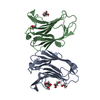 7n4oC 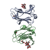 7n57C 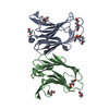 7n6cC 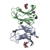 7n8dC 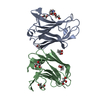 7n8gC 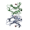 7n96C  4bkzS S: Starting model for refinement C: citing same article ( |
|---|---|
| Similar structure data |
-Links
 PDBj
PDBj
- Assembly
| Deposited unit | 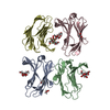
| ||||||||||||||||||||||||||||||||||||||||||||||||||||||||||||||||||||||||||||||
|---|---|---|---|---|---|---|---|---|---|---|---|---|---|---|---|---|---|---|---|---|---|---|---|---|---|---|---|---|---|---|---|---|---|---|---|---|---|---|---|---|---|---|---|---|---|---|---|---|---|---|---|---|---|---|---|---|---|---|---|---|---|---|---|---|---|---|---|---|---|---|---|---|---|---|---|---|---|---|---|
| 1 | 
| ||||||||||||||||||||||||||||||||||||||||||||||||||||||||||||||||||||||||||||||
| 2 | 
| ||||||||||||||||||||||||||||||||||||||||||||||||||||||||||||||||||||||||||||||
| Unit cell |
| ||||||||||||||||||||||||||||||||||||||||||||||||||||||||||||||||||||||||||||||
| Noncrystallographic symmetry (NCS) | NCS domain:
NCS domain segments: Ens-ID: 1
|