 Movie
Movie Controller

[English] 日本語
 Yorodumi
- PDB-7raq: Crystal structure of CV3-25 Fab bound to SARS-CoV-2 spike stem he... -
+ Open data

- Basic information
| Entry | Database: PDB / ID: 7raq | ||||||
|---|---|---|---|---|---|---|---|
| Title | Crystal structure of CV3-25 Fab bound to SARS-CoV-2 spike stem helix peptide | ||||||
 Components Components |
| ||||||
 Keywords Keywords | IMMUNE SYSTEM / Antibody / Fab / SARS-CoV-2 | ||||||
| Function / homology |  Function and homology information Function and homology informationsymbiont-mediated disruption of host tissue / Maturation of spike protein / Translation of Structural Proteins / Virion Assembly and Release / host cell surface / host extracellular region / symbiont-mediated-mediated suppression of host tetherin activity / Induction of Cell-Cell Fusion / structural constituent of virion / positive regulation of viral entry into host cell ...symbiont-mediated disruption of host tissue / Maturation of spike protein / Translation of Structural Proteins / Virion Assembly and Release / host cell surface / host extracellular region / symbiont-mediated-mediated suppression of host tetherin activity / Induction of Cell-Cell Fusion / structural constituent of virion / positive regulation of viral entry into host cell / membrane fusion / host cell endoplasmic reticulum-Golgi intermediate compartment membrane / Attachment and Entry / entry receptor-mediated virion attachment to host cell / receptor-mediated virion attachment to host cell / host cell surface receptor binding / symbiont-mediated suppression of host innate immune response / endocytosis involved in viral entry into host cell / receptor ligand activity / fusion of virus membrane with host plasma membrane / fusion of virus membrane with host endosome membrane / viral envelope / symbiont entry into host cell / virion attachment to host cell / host cell plasma membrane / SARS-CoV-2 activates/modulates innate and adaptive immune responses / virion membrane / membrane / identical protein binding / plasma membrane Similarity search - Function | ||||||
| Biological species |  Homo sapiens (human) Homo sapiens (human)  Severe acute respiratory syndrome coronavirus 2 Severe acute respiratory syndrome coronavirus 2 | ||||||
| Method |  X-RAY DIFFRACTION / SYNCHROTRON / MOLECULAR REPLACEMENT / Resolution: 1.74 Å X-RAY DIFFRACTION / SYNCHROTRON / MOLECULAR REPLACEMENT / Resolution: 1.74 Å | ||||||
 Authors Authors | Hurlburt, N.K. / Pancera, M. | ||||||
 Citation Citation | Journal: Commun Biol / Year: 2022 Title: Structural definition of a pan-sarbecovirus neutralizing epitope on the spike S2 subunit. Authors: Nicholas K Hurlburt / Leah J Homad / Irika Sinha / Madeleine F Jennewein / Anna J MacCamy / Yu-Hsin Wan / Jim Boonyaratanakornkit / Anton M Sholukh / Abigail M Jackson / Panpan Zhou / Dennis ...Authors: Nicholas K Hurlburt / Leah J Homad / Irika Sinha / Madeleine F Jennewein / Anna J MacCamy / Yu-Hsin Wan / Jim Boonyaratanakornkit / Anton M Sholukh / Abigail M Jackson / Panpan Zhou / Dennis R Burton / Raiees Andrabi / Gabriel Ozorowski / Andrew B Ward / Leonidas Stamatatos / Marie Pancera / Andrew T McGuire /  Abstract: Three betacoronaviruses have crossed the species barrier and established human-to-human transmission causing significant morbidity and mortality in the past 20 years. The most current and widespread ...Three betacoronaviruses have crossed the species barrier and established human-to-human transmission causing significant morbidity and mortality in the past 20 years. The most current and widespread of these is SARS-CoV-2. The identification of CoVs with zoonotic potential in animal reservoirs suggests that additional outbreaks could occur. Monoclonal antibodies targeting conserved neutralizing epitopes on diverse CoVs can form the basis for prophylaxis and therapeutic treatments and enable the design of vaccines aimed at providing pan-CoV protection. We previously identified a neutralizing monoclonal antibody, CV3-25 that binds to the SARS-CoV-2 spike, neutralizes the SARS-CoV-2 Beta variant comparably to the ancestral Wuhan Hu-1 strain, cross neutralizes SARS-CoV-1 and binds to recombinant proteins derived from the spike-ectodomains of HCoV-OC43 and HCoV-HKU1. Here, we show that the neutralizing activity of CV3-25 is maintained against the Alpha, Delta, Gamma and Omicron variants of concern as well as a SARS-CoV-like bat coronavirus with zoonotic potential by binding to a conserved linear peptide in the stem-helix region. Negative stain electron microscopy and a 1.74 Å crystal structure of a CV3-25/peptide complex demonstrates that CV3-25 binds to the base of the stem helix at the HR2 boundary to an epitope that is distinct from other stem-helix directed neutralizing mAbs. | ||||||
| History |
|
- Structure visualization
| Structure viewer | Molecule: MolmilJmol/JSmol |
|---|
- Downloads & links
-Download
| PDBx/mmCIF format | 7raq.cif.gz | 197.1 KB | Display | PDBx/mmCIF format |
|---|---|---|---|---|
| PDB format | pdb7raq.ent.gz | 154.7 KB | Display | PDB format |
| PDBx/mmJSON format | 7raq.json.gz | Tree view | PDBx/mmJSON format | |
| Others |  Other downloads Other downloads |
-Validation report
| Arichive directory | https://data.pdbj.org/pub/pdb/validation_reports/ra/7raqftp://data.pdbj.org/pub/pdb/validation_reports/ra/7raq | HTTPS FTP |
|---|
-Related structure data
| Related structure data |  5mvzS S: Starting model for refinement C: citing same article ( |
|---|---|
| Similar structure data |

-Links
 PDBj
PDBj


- Assembly
| Deposited unit | 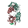
| ||||||||||
|---|---|---|---|---|---|---|---|---|---|---|---|
| 1 |
| ||||||||||
| Unit cell |
| ||||||||||
| Components on special symmetry positions |
|
-Components
-Protein/peptide , 1 types, 1 molecules P
| #3: Protein/peptide | Mass: 2239.437 Da / Num. of mol.: 1 / Source method: obtained synthetically Source: (synth.) Severe acute respiratory syndrome coronavirus 2References: UniProt: P0DTC2 |
|---|
-Antibody , 2 types, 2 molecules HL
| #1: Antibody | Mass: 25026.203 Da / Num. of mol.: 1 Source method: isolated from a genetically manipulated source Source: (gene. exp.) Homo sapiens (human) / Cell line (production host): HEK 293E / Production host: Homo sapiens (human) |
|---|---|
| #2: Antibody | Mass: 23218.695 Da / Num. of mol.: 1 Source method: isolated from a genetically manipulated source Source: (gene. exp.) Homo sapiens (human) / Cell line (production host): HEK 293E / Production host: Homo sapiens (human) |
-Non-polymers , 3 types, 303 molecules 


| #4: Chemical | ChemComp-GOL /  Mass: 92.094 Da / Num. of mol.: 7 / Source method: obtained synthetically / Formula: C3H8O3 Mass: 92.094 Da / Num. of mol.: 7 / Source method: obtained synthetically / Formula: C3H8O3#5: Chemical |  Mass: 96.063 Da / Num. of mol.: 2 / Source method: obtained synthetically / Formula: SO4 Mass: 96.063 Da / Num. of mol.: 2 / Source method: obtained synthetically / Formula: SO4#6: Water | ChemComp-HOH / | Mass: 18.015 Da / Num. of mol.: 294 / Source method: isolated from a natural source / Formula: H2O |
|---|
-Details
| Has ligand of interest | N |
|---|---|
| Has protein modification | Y |
-Experimental details
-Experiment
| Experiment | Method: X-RAY DIFFRACTION / Number of used crystals: 1 |
|---|
- Sample preparation
| Crystal | Density Matthews: 2.96 Å3/Da / Density % sol: 58.42 % |
|---|---|
| Crystal grow | Temperature: 293 K / Method: vapor diffusion, hanging drop Details: 0.1M Na Acetate:HCl, pH 4.5, 2.0M (NH4)2SO4, 0.01M SrCl |
-Data collection
| Diffraction | Mean temperature: 100 K / Serial crystal experiment: N | ||||||||||||||||||||||||||||||
|---|---|---|---|---|---|---|---|---|---|---|---|---|---|---|---|---|---|---|---|---|---|---|---|---|---|---|---|---|---|---|---|
| Diffraction source | Source: SYNCHROTRON / Site: ALS / Beamline: 5.0.2 / Wavelength: 1.0092 Å | ||||||||||||||||||||||||||||||
| Detector | Type: DECTRIS PILATUS 6M / Detector: PIXEL / Date: Jun 10, 2021 | ||||||||||||||||||||||||||||||
| Radiation | Protocol: SINGLE WAVELENGTH / Monochromatic (M) / Laue (L): M / Scattering type: x-ray | ||||||||||||||||||||||||||||||
| Radiation wavelength | Wavelength: 1.0092 Å / Relative weight: 1 | ||||||||||||||||||||||||||||||
| Reflection | Resolution: 1.74→49.01 Å / Num. obs: 63310 / % possible obs: 100 % / Redundancy: 1.9 % / CC1/2: 0.999 / Rmerge(I) obs: 0.025 / Rpim(I) all: 0.025 / Rrim(I) all: 0.035 / Net I/σ(I): 21.7 | ||||||||||||||||||||||||||||||
| Reflection shell | Diffraction-ID: 1
|
- Processing
| Software |
| ||||||||||||||||||||||||||||||||||||||||||||||||||||||||||||||||||||||||||||||||||||||||||||||||||||
|---|---|---|---|---|---|---|---|---|---|---|---|---|---|---|---|---|---|---|---|---|---|---|---|---|---|---|---|---|---|---|---|---|---|---|---|---|---|---|---|---|---|---|---|---|---|---|---|---|---|---|---|---|---|---|---|---|---|---|---|---|---|---|---|---|---|---|---|---|---|---|---|---|---|---|---|---|---|---|---|---|---|---|---|---|---|---|---|---|---|---|---|---|---|---|---|---|---|---|---|---|---|
| Refinement | Method to determine structure: MOLECULAR REPLACEMENT Starting model: 5MVZ Resolution: 1.74→49.01 Å / Cor.coef. Fo:Fc: 0.967 / Cor.coef. Fo:Fc free: 0.958 / SU B: 4.274 / SU ML: 0.068 / Cross valid method: THROUGHOUT / ESU R: 0.094 / ESU R Free: 0.091 / Stereochemistry target values: MAXIMUM LIKELIHOOD Details: U VALUES : WITH TLS ADDED HYDROGENS HAVE BEEN ADDED IN THE RIDING POSITIONS U VALUES : RESIDUAL ONLY
| ||||||||||||||||||||||||||||||||||||||||||||||||||||||||||||||||||||||||||||||||||||||||||||||||||||
| Solvent computation | Ion probe radii: 0.7 Å / Shrinkage radii: 0.7 Å / VDW probe radii: 1.3 Å / Solvent model: MASK | ||||||||||||||||||||||||||||||||||||||||||||||||||||||||||||||||||||||||||||||||||||||||||||||||||||
| Displacement parameters | Biso max: 93.51 Å2 / Biso mean: 36.109 Å2 / Biso min: 19.21 Å2
| ||||||||||||||||||||||||||||||||||||||||||||||||||||||||||||||||||||||||||||||||||||||||||||||||||||
| Refinement step | Cycle: final / Resolution: 1.74→49.01 Å
| ||||||||||||||||||||||||||||||||||||||||||||||||||||||||||||||||||||||||||||||||||||||||||||||||||||
| Refine LS restraints |
| ||||||||||||||||||||||||||||||||||||||||||||||||||||||||||||||||||||||||||||||||||||||||||||||||||||
| LS refinement shell | Resolution: 1.74→1.785 Å / Rfactor Rfree error: 0 / Total num. of bins used: 20
| ||||||||||||||||||||||||||||||||||||||||||||||||||||||||||||||||||||||||||||||||||||||||||||||||||||
| Refinement TLS params. | Method: refined / Refine-ID: X-RAY DIFFRACTION
| ||||||||||||||||||||||||||||||||||||||||||||||||||||||||||||||||||||||||||||||||||||||||||||||||||||
| Refinement TLS group |
|
