 Movie
Movie Controller

[English] 日本語
 Yorodumi
- PDB-7r6b: Crystal structure of mutant R43D/L124D/R125A/C273S of L-Asparagin... -
+ Open data

- Basic information
| Entry | Database: PDB / ID: 7r6b | ||||||
|---|---|---|---|---|---|---|---|
| Title | Crystal structure of mutant R43D/L124D/R125A/C273S of L-Asparaginase I from Yersinia pestis | ||||||
 Components Components | L-asparaginase I | ||||||
 Keywords Keywords | HYDROLASE / L-ASPARAGINASE | ||||||
| Function / homology |  Function and homology information Function and homology informationasparaginase / asparaginase activity / amino acid metabolic process / metal ion binding / cytosol Similarity search - Function | ||||||
| Biological species |   Yersinia pestis (bacteria) Yersinia pestis (bacteria) | ||||||
| Method |  X-RAY DIFFRACTION / SYNCHROTRON / MOLECULAR REPLACEMENT / molecular replacement / Resolution: 2.03 Å X-RAY DIFFRACTION / SYNCHROTRON / MOLECULAR REPLACEMENT / molecular replacement / Resolution: 2.03 Å | ||||||
 Authors Authors | Strzelczyk, P. / Wlodawer, A. / Lubkowski, J. | ||||||
| Funding support |  United States, 1items United States, 1items
| ||||||
 Citation Citation | Journal: Febs J. / Year: 2023 Title: The dimeric form of bacterial l-asparaginase YpAI is fully active. Authors: Strzelczyk, P. / Zhang, D. / Alexandratos, J. / Piszczek, G. / Wlodawer, A. / Lubkowski, J. | ||||||
| History |
|
- Structure visualization
| Structure viewer | Molecule: MolmilJmol/JSmol |
|---|
- Downloads & links
-Download
| PDBx/mmCIF format | 7r6b.cif.gz | 140.9 KB | Display | PDBx/mmCIF format |
|---|---|---|---|---|
| PDB format | pdb7r6b.ent.gz | 107.2 KB | Display | PDB format |
| PDBx/mmJSON format | 7r6b.json.gz | Tree view | PDBx/mmJSON format | |
| Others |  Other downloads Other downloads |
-Validation report
| Summary document | 7r6b_validation.pdf.gz | 462.1 KB | Display | wwPDB validaton report |
|---|---|---|---|---|
| Full document | 7r6b_full_validation.pdf.gz | 477.3 KB | Display | |
| Data in XML | 7r6b_validation.xml.gz | 29.2 KB | Display | |
| Data in CIF | 7r6b_validation.cif.gz | 41.9 KB | Display | |
| Arichive directory | https://data.pdbj.org/pub/pdb/validation_reports/r6/7r6bftp://data.pdbj.org/pub/pdb/validation_reports/r6/7r6b | HTTPS FTP |
-Related structure data
| Related structure data |  7r69C  7r6aC 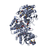 3ntxS S: Starting model for refinement C: citing same article ( |
|---|---|
| Similar structure data |
-Links
 PDBj
PDBj- Assembly
| Deposited unit | 
| ||||||||||||||||||
|---|---|---|---|---|---|---|---|---|---|---|---|---|---|---|---|---|---|---|---|
| 1 |
| ||||||||||||||||||
| Unit cell |
| ||||||||||||||||||
| Noncrystallographic symmetry (NCS) | NCS domain:
NCS domain segments: Component-ID: _ / Ens-ID: 1 / Beg auth comp-ID: LYS / Beg label comp-ID: LYS / End auth comp-ID: ASP / End label comp-ID: ASP / Refine code: _ / Auth seq-ID: 3 - 336 / Label seq-ID: 3 - 336
|
-Components
| #1: Protein | Mass: 36690.309 Da / Num. of mol.: 2 / Mutation: R43D, L124D, R125A, C273S Source method: isolated from a genetically manipulated source Source: (gene. exp.) Yersinia pestis (bacteria) / Gene: ansA, YPO2161 / Plasmid: pET22b(+) / Cell (production host): Bacteria / Production host: #2: Chemical | ChemComp-EDO /   Mass: 62.068 Da / Num. of mol.: 10 / Source method: obtained synthetically / Formula: C2H6O2 Mass: 62.068 Da / Num. of mol.: 10 / Source method: obtained synthetically / Formula: C2H6O2#3: Chemical |   Mass: 40.078 Da / Num. of mol.: 2 / Source method: obtained synthetically / Formula: Ca Mass: 40.078 Da / Num. of mol.: 2 / Source method: obtained synthetically / Formula: Ca#4: Water | ChemComp-HOH / |  Mass: 18.015 Da / Num. of mol.: 383 / Source method: isolated from a natural source / Formula: H2O Mass: 18.015 Da / Num. of mol.: 383 / Source method: isolated from a natural source / Formula: H2OHas ligand of interest | N | |
|---|
-Experimental details
-Experiment
| Experiment | Method: X-RAY DIFFRACTION / Number of used crystals: 1 |
|---|
- Sample preparation
| Crystal | Density Matthews: 2.52 Å3/Da / Density % sol: 51.28 % |
|---|---|
| Crystal grow | Temperature: 293 K / Method: vapor diffusion / pH: 8 Details: 0.2 M Calcium acetate, 10% (w/v) PEG 8000, 0.1 M Imidazole/ Hydrochloric acid, pH 8.0 |
-Data collection
| Diffraction | Mean temperature: 100 K / Serial crystal experiment: N |
|---|---|
| Diffraction source | Source: SYNCHROTRON / Site: APS / Beamline: 22-ID / Wavelength: 1 Å |
| Detector | Type: DECTRIS EIGER X 16M / Detector: PIXEL / Date: Mar 6, 2020 |
| Radiation | Monochromator: Double crystal - liquid nitrogen cooled Si(111) Protocol: SINGLE WAVELENGTH / Monochromatic (M) / Laue (L): M / Scattering type: x-ray |
| Radiation wavelength | Wavelength: 1 Å / Relative weight: 1 |
| Reflection | Resolution: 2.03→50 Å / Num. obs: 47794 / % possible obs: 98.7 % / Redundancy: 5.7 % / CC1/2: 0.997 / Rmerge(I) obs: 0.092 / Rpim(I) all: 0.042 / Net I/σ(I): 22 |
| Reflection shell | Resolution: 2.03→2.07 Å / Redundancy: 4.6 % / Rmerge(I) obs: 0.92 / Mean I/σ(I) obs: 2.1 / Num. unique obs: 2370 / CC1/2: 0.719 / Rpim(I) all: 0.47 / % possible all: 99.5 |
-Phasing
| Phasing | Method: molecular replacement |
|---|
- Processing
| Software |
| ||||||||||||||||||||||||||||||||||||||||||||||||||||||||||||
|---|---|---|---|---|---|---|---|---|---|---|---|---|---|---|---|---|---|---|---|---|---|---|---|---|---|---|---|---|---|---|---|---|---|---|---|---|---|---|---|---|---|---|---|---|---|---|---|---|---|---|---|---|---|---|---|---|---|---|---|---|---|
| Refinement | Method to determine structure: MOLECULAR REPLACEMENT Starting model: 3NTX Resolution: 2.03→48.85 Å / Cor.coef. Fo:Fc: 0.96 / Cor.coef. Fo:Fc free: 0.939 / SU B: 4.234 / SU ML: 0.114 / Cross valid method: THROUGHOUT / σ(F): 0 / ESU R: 0.164 / ESU R Free: 0.155 / Stereochemistry target values: MAXIMUM LIKELIHOOD Details: HYDROGENS HAVE BEEN ADDED IN THE RIDING POSITIONS U VALUES : REFINED INDIVIDUALLY
| ||||||||||||||||||||||||||||||||||||||||||||||||||||||||||||
| Solvent computation | Ion probe radii: 0.8 Å / Shrinkage radii: 0.8 Å / VDW probe radii: 1.2 Å / Solvent model: MASK | ||||||||||||||||||||||||||||||||||||||||||||||||||||||||||||
| Displacement parameters | Biso max: 113.19 Å2 / Biso mean: 31.099 Å2 / Biso min: 11.55 Å2
| ||||||||||||||||||||||||||||||||||||||||||||||||||||||||||||
| Refinement step | Cycle: final / Resolution: 2.03→48.85 Å
| ||||||||||||||||||||||||||||||||||||||||||||||||||||||||||||
| Refine LS restraints |
| ||||||||||||||||||||||||||||||||||||||||||||||||||||||||||||
| Refine LS restraints NCS | Ens-ID: 1 / Number: 8892 / Refine-ID: X-RAY DIFFRACTION / Type: interatomic distance / Rms dev position: 0.13 Å / Weight position: 0.05
| ||||||||||||||||||||||||||||||||||||||||||||||||||||||||||||
| LS refinement shell | Resolution: 2.03→2.08 Å / Rfactor Rfree error: 0
|
