 Movie
Movie Controller


+ Open data

- Basic information
| Entry | Database: PDB / ID: 7r25 | |||||||||
|---|---|---|---|---|---|---|---|---|---|---|
| Title | Bacillus pumilus Lipase A | |||||||||
 Components Components | Lipase | |||||||||
 Keywords Keywords | HYDROLASE / lipase / esterase / apo | |||||||||
| Function / homology | Lipase EstA/Esterase EstB / Lipase (class 2) / lipase activity / lipid catabolic process / Alpha/Beta hydrolase fold / CITRIC ACID / PHOSPHATE ION / PROPANOIC ACID / Lipase Function and homology information Function and homology information | |||||||||
| Biological species |  | |||||||||
| Method |  X-RAY DIFFRACTION / SYNCHROTRON / MOLECULAR REPLACEMENT / Resolution: 0.87 Å X-RAY DIFFRACTION / SYNCHROTRON / MOLECULAR REPLACEMENT / Resolution: 0.87 Å | |||||||||
 Authors Authors | Lund, B.A. | |||||||||
| Funding support |  Norway, 2items Norway, 2items
| |||||||||
 Citation Citation | Journal: Biochemistry / Year: 2022 Title: Structure and Mechanism of a Cold-Adapted Bacterial Lipase Authors: Lund, B.A. / Svalberg, L. / Purg, M. / Chukwu, G. / Widersten, M. / Isaksen, G.V. / Brandsdal, B.O. / Aqvist, J. | |||||||||
| History |
|
- Structure visualization
| Structure viewer | Molecule: MolmilJmol/JSmol |
|---|
- Downloads & links
-Download
| PDBx/mmCIF format | 7r25.cif.gz | 149.8 KB | Display | PDBx/mmCIF format |
|---|---|---|---|---|
| PDB format | pdb7r25.ent.gz | 103.2 KB | Display | PDB format |
| PDBx/mmJSON format | 7r25.json.gz | Tree view | PDBx/mmJSON format | |
| Others |  Other downloads Other downloads |
-Validation report
| Arichive directory | https://data.pdbj.org/pub/pdb/validation_reports/r2/7r25ftp://data.pdbj.org/pub/pdb/validation_reports/r2/7r25 | HTTPS FTP |
|---|
-Related structure data
| Related structure data | 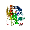 7r1kC 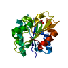 1ispS S: Starting model for refinement C: citing same article ( |
|---|---|
| Similar structure data |
-Links
 PDBj
PDBj
- Assembly
| Deposited unit | 
| ||||||||||||
|---|---|---|---|---|---|---|---|---|---|---|---|---|---|
| 1 |
| ||||||||||||
| Unit cell |
|
-Components
| #1: Protein | Mass: 20483.234 Da / Num. of mol.: 1 Source method: isolated from a genetically manipulated source Source: (gene. exp.) |
|---|---|
| #2: Chemical | ChemComp-CIT /   Mass: 192.124 Da / Num. of mol.: 1 / Source method: obtained synthetically / Formula: C6H8O7 Mass: 192.124 Da / Num. of mol.: 1 / Source method: obtained synthetically / Formula: C6H8O7 |
| #3: Chemical | ChemComp-PO4 /   Mass: 94.971 Da / Num. of mol.: 1 / Source method: obtained synthetically / Formula: PO4 Mass: 94.971 Da / Num. of mol.: 1 / Source method: obtained synthetically / Formula: PO4 |
| #4: Chemical | ChemComp-PPI / 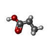  Mass: 74.079 Da / Num. of mol.: 1 / Source method: obtained synthetically / Formula: C3H6O2 Mass: 74.079 Da / Num. of mol.: 1 / Source method: obtained synthetically / Formula: C3H6O2 |
| #5: Water | ChemComp-HOH /  Mass: 18.015 Da / Num. of mol.: 273 / Source method: isolated from a natural source / Formula: H2O Mass: 18.015 Da / Num. of mol.: 273 / Source method: isolated from a natural source / Formula: H2O |
| Has ligand of interest | N |
-Experimental details
-Experiment
| Experiment | Method: X-RAY DIFFRACTION / Number of used crystals: 1 |
|---|
- Sample preparation
| Crystal | Density Matthews: 1.92 Å3/Da / Density % sol: 35.88 % |
|---|---|
| Crystal grow | Temperature: 277 K / Method: vapor diffusion, hanging drop / pH: 6.5 Details: 0.1 M MES pH 6.5 18-22 % PEG 4000 0.1-0.5 M lithium citrate |
-Data collection
| Diffraction | Mean temperature: 100 K / Serial crystal experiment: N |
|---|---|
| Diffraction source | Source: SYNCHROTRON / Site: BESSY  / Beamline: 14.1 / Wavelength: 0.9184 Å / Beamline: 14.1 / Wavelength: 0.9184 Å |
| Detector | Type: DECTRIS PILATUS 6M / Detector: PIXEL / Date: Mar 6, 2020 |
| Radiation | Protocol: SINGLE WAVELENGTH / Monochromatic (M) / Laue (L): M / Scattering type: x-ray |
| Radiation wavelength | Wavelength: 0.9184 Å / Relative weight: 1 |
| Reflection | Resolution: 0.87→35.07 Å / Num. obs: 103387 / % possible obs: 81.78 % / Redundancy: 6.1 % / Biso Wilson estimate: 7.03 Å2 / CC1/2: 1 / Rmerge(I) obs: 0.028 / Net I/σ(I): 30.45 |
| Reflection shell | Resolution: 0.87→0.9013 Å / Rmerge(I) obs: 0.4447 / Mean I/σ(I) obs: 2.15 / Num. unique obs: 1919 / CC1/2: 0.786 / % possible all: 3.1 |
- Processing
| Software |
| ||||||||||||||||||||||||||||||||||||||||||||||||||||||||||||||||||||||||||||||||||||||||||||||||||||||||||||||||
|---|---|---|---|---|---|---|---|---|---|---|---|---|---|---|---|---|---|---|---|---|---|---|---|---|---|---|---|---|---|---|---|---|---|---|---|---|---|---|---|---|---|---|---|---|---|---|---|---|---|---|---|---|---|---|---|---|---|---|---|---|---|---|---|---|---|---|---|---|---|---|---|---|---|---|---|---|---|---|---|---|---|---|---|---|---|---|---|---|---|---|---|---|---|---|---|---|---|---|---|---|---|---|---|---|---|---|---|---|---|---|---|---|---|
| Refinement | Method to determine structure: MOLECULAR REPLACEMENT Starting model: 1isp Resolution: 0.87→35.07 Å / SU ML: 0.0492 / Cross valid method: FREE R-VALUE / σ(F): 1.38 / Phase error: 13.8355 Stereochemistry target values: GeoStd + Monomer Library + CDL v1.2
| ||||||||||||||||||||||||||||||||||||||||||||||||||||||||||||||||||||||||||||||||||||||||||||||||||||||||||||||||
| Solvent computation | Shrinkage radii: 1.3 Å / VDW probe radii: 1.4 Å / Solvent model: FLAT BULK SOLVENT MODEL | ||||||||||||||||||||||||||||||||||||||||||||||||||||||||||||||||||||||||||||||||||||||||||||||||||||||||||||||||
| Displacement parameters | Biso mean: 12.66 Å2 | ||||||||||||||||||||||||||||||||||||||||||||||||||||||||||||||||||||||||||||||||||||||||||||||||||||||||||||||||
| Refinement step | Cycle: LAST / Resolution: 0.87→35.07 Å
| ||||||||||||||||||||||||||||||||||||||||||||||||||||||||||||||||||||||||||||||||||||||||||||||||||||||||||||||||
| Refine LS restraints |
| ||||||||||||||||||||||||||||||||||||||||||||||||||||||||||||||||||||||||||||||||||||||||||||||||||||||||||||||||
| LS refinement shell |
|
