 Movie
Movie Controller


+ Open data

- Basic information
| Entry | Database: PDB / ID: 7r20 | ||||||
|---|---|---|---|---|---|---|---|
| Title | Anti-Arc nanobody E5 | ||||||
 Components Components | Anti-Arc nanobody E5 | ||||||
 Keywords Keywords | IMMUNE SYSTEM / Nanobody / VHH | ||||||
| Function / homology | Immunoglobulins / Immunoglobulin-like / Sandwich / Mainly Beta Function and homology information Function and homology information | ||||||
| Biological species |  | ||||||
| Method |  X-RAY DIFFRACTION / SYNCHROTRON / MOLECULAR REPLACEMENT / Resolution: 1.42 Å X-RAY DIFFRACTION / SYNCHROTRON / MOLECULAR REPLACEMENT / Resolution: 1.42 Å | ||||||
 Authors Authors | Markusson, S. / Kursula, P. | ||||||
| Funding support |  Norway, 1items Norway, 1items
| ||||||
 Citation Citation | Journal: Plos One / Year: 2022 Title: High-affinity anti-Arc nanobodies provide tools for structural and functional studies. Authors: Markusson, S. / Hallin, E.I. / Bustad, H.J. / Raasakka, A. / Xu, J. / Muruganandam, G. / Loris, R. / Martinez, A. / Bramham, C.R. / Kursula, P. | ||||||
| History |
|
- Structure visualization
| Structure viewer | Molecule: MolmilJmol/JSmol |
|---|
- Downloads & links
-Download
| PDBx/mmCIF format | 7r20.cif.gz | 101.7 KB | Display | PDBx/mmCIF format |
|---|---|---|---|---|
| PDB format | pdb7r20.ent.gz | 65.4 KB | Display | PDB format |
| PDBx/mmJSON format | 7r20.json.gz | Tree view | PDBx/mmJSON format | |
| Others |  Other downloads Other downloads |
-Validation report
| Arichive directory | https://data.pdbj.org/pub/pdb/validation_reports/r2/7r20ftp://data.pdbj.org/pub/pdb/validation_reports/r2/7r20 | HTTPS FTP |
|---|
-Related structure data
| Related structure data |  7r1zC  5hvgS S: Starting model for refinement C: citing same article ( |
|---|---|
| Similar structure data |
-Links
 PDBj
PDBj


- Assembly
| Deposited unit | 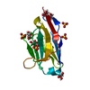
| ||||||||||||
|---|---|---|---|---|---|---|---|---|---|---|---|---|---|
| 1 |
| ||||||||||||
| Unit cell |
|
-Components
| #1: Antibody | Mass: 12767.269 Da / Num. of mol.: 1 Source method: isolated from a genetically manipulated source Source: (gene. exp.)  | ||||||||
|---|---|---|---|---|---|---|---|---|---|
| #2: Chemical | ChemComp-GOL /   Mass: 92.094 Da / Num. of mol.: 6 / Source method: obtained synthetically / Formula: C3H8O3 Mass: 92.094 Da / Num. of mol.: 6 / Source method: obtained synthetically / Formula: C3H8O3#3: Chemical | ChemComp-SO4 /   Mass: 96.063 Da / Num. of mol.: 4 / Source method: obtained synthetically / Formula: SO4 Mass: 96.063 Da / Num. of mol.: 4 / Source method: obtained synthetically / Formula: SO4#4: Water | ChemComp-HOH / |  Mass: 18.015 Da / Num. of mol.: 47 / Source method: isolated from a natural source / Formula: H2O Mass: 18.015 Da / Num. of mol.: 47 / Source method: isolated from a natural source / Formula: H2OHas ligand of interest | N | Has protein modification | Y | |
-Experimental details
-Experiment
| Experiment | Method: X-RAY DIFFRACTION / Number of used crystals: 1 |
|---|
- Sample preparation
| Crystal | Density Matthews: 2.02 Å3/Da / Density % sol: 39.3 % |
|---|---|
| Crystal grow | Temperature: 293 K / Method: vapor diffusion, sitting drop / pH: 7.5 Details: 1.26 M (NH4)2SO4, 0.1 M LiSO4 and 0.1 M Tris-HCl pH 7.5 |
-Data collection
| Diffraction | Mean temperature: 100 K / Serial crystal experiment: N |
|---|---|
| Diffraction source | Source: SYNCHROTRON / Site: Diamond  / Beamline: I03 / Wavelength: 0.976 Å / Beamline: I03 / Wavelength: 0.976 Å |
| Detector | Type: DECTRIS EIGER2 X 16M / Detector: PIXEL / Date: Feb 21, 2021 |
| Radiation | Protocol: SINGLE WAVELENGTH / Monochromatic (M) / Laue (L): M / Scattering type: x-ray |
| Radiation wavelength | Wavelength: 0.976 Å / Relative weight: 1 |
| Reflection | Resolution: 1.42→41.98 Å / Num. obs: 201623 / % possible obs: 99.9 % / Redundancy: 9.2 % / Biso Wilson estimate: 24.51 Å2 / CC1/2: 0.999 / Rrim(I) all: 0.108 / Net I/σ(I): 9.5 |
| Reflection shell | Resolution: 1.42→1.46 Å / Redundancy: 9.2 % / Mean I/σ(I) obs: 0.3 / Num. unique obs: 21801 / CC1/2: 0.333 / Rrim(I) all: 5.687 / % possible all: 99.8 |
- Processing
| Software |
| |||||||||||||||||||||||||||||||||||||||||||||||||||||||||||||||||||||||||||||||||||||||||||||||||||||||||
|---|---|---|---|---|---|---|---|---|---|---|---|---|---|---|---|---|---|---|---|---|---|---|---|---|---|---|---|---|---|---|---|---|---|---|---|---|---|---|---|---|---|---|---|---|---|---|---|---|---|---|---|---|---|---|---|---|---|---|---|---|---|---|---|---|---|---|---|---|---|---|---|---|---|---|---|---|---|---|---|---|---|---|---|---|---|---|---|---|---|---|---|---|---|---|---|---|---|---|---|---|---|---|---|---|---|---|
| Refinement | Method to determine structure: MOLECULAR REPLACEMENT Starting model: 5HVG Resolution: 1.42→41.98 Å / SU ML: 0.2566 / Cross valid method: FREE R-VALUE / σ(F): 1.33 / Phase error: 33.7126 Stereochemistry target values: GeoStd + Monomer Library + CDL v1.2
| |||||||||||||||||||||||||||||||||||||||||||||||||||||||||||||||||||||||||||||||||||||||||||||||||||||||||
| Solvent computation | Shrinkage radii: 0.9 Å / VDW probe radii: 1.11 Å / Solvent model: FLAT BULK SOLVENT MODEL | |||||||||||||||||||||||||||||||||||||||||||||||||||||||||||||||||||||||||||||||||||||||||||||||||||||||||
| Displacement parameters | Biso mean: 35.08 Å2 | |||||||||||||||||||||||||||||||||||||||||||||||||||||||||||||||||||||||||||||||||||||||||||||||||||||||||
| Refinement step | Cycle: LAST / Resolution: 1.42→41.98 Å
| |||||||||||||||||||||||||||||||||||||||||||||||||||||||||||||||||||||||||||||||||||||||||||||||||||||||||
| Refine LS restraints |
| |||||||||||||||||||||||||||||||||||||||||||||||||||||||||||||||||||||||||||||||||||||||||||||||||||||||||
| LS refinement shell |
|
