 Movie
Movie Controller

[English] 日本語
 Yorodumi
- PDB-7r1v: Crystal structure of E.coli BamA beta-barrel in complex with dyno... -
+ Open data

- Basic information
| Entry | Database: PDB / ID: 7r1v | ||||||
|---|---|---|---|---|---|---|---|
| Title | Crystal structure of E.coli BamA beta-barrel in complex with dynobactin A | ||||||
 Components Components |
| ||||||
 Keywords Keywords | MEMBRANE PROTEIN / Beta-Barrel / outer membrane / protein insertion / protein folding / protein maturation / antibiotic / natural product / cyclized peptide | ||||||
| Function / homology |  Function and homology information Function and homology informationBam protein complex / Gram-negative-bacterium-type cell outer membrane assembly / protein insertion into membrane Similarity search - Function | ||||||
| Biological species |  Photorhabdus australis (bacteria) Photorhabdus australis (bacteria) | ||||||
| Method |  X-RAY DIFFRACTION / SYNCHROTRON / MOLECULAR REPLACEMENT / Resolution: 2.5 Å X-RAY DIFFRACTION / SYNCHROTRON / MOLECULAR REPLACEMENT / Resolution: 2.5 Å | ||||||
 Authors Authors | Jakob, R.P. / Hiller, S. / Maier, T. | ||||||
| Funding support |  Switzerland, 1items Switzerland, 1items
| ||||||
 Citation Citation | Journal: Nat Microbiol / Year: 2022 Title: Computational identification of a systemic antibiotic for gram-negative bacteria. Authors: Ryan D Miller / Akira Iinishi / Seyed Majed Modaresi / Byung-Kuk Yoo / Thomas D Curtis / Patrick J Lariviere / Libang Liang / Sangkeun Son / Samantha Nicolau / Rachel Bargabos / Madeleine ...Authors: Ryan D Miller / Akira Iinishi / Seyed Majed Modaresi / Byung-Kuk Yoo / Thomas D Curtis / Patrick J Lariviere / Libang Liang / Sangkeun Son / Samantha Nicolau / Rachel Bargabos / Madeleine Morrissette / Michael F Gates / Norman Pitt / Roman P Jakob / Parthasarathi Rath / Timm Maier / Andrey G Malyutin / Jens T Kaiser / Samantha Niles / Blake Karavas / Meghan Ghiglieri / Sarah E J Bowman / Douglas C Rees / Sebastian Hiller / Kim Lewis /  Abstract: Discovery of antibiotics acting against Gram-negative species is uniquely challenging due to their restrictive penetration barrier. BamA, which inserts proteins into the outer membrane, is an ...Discovery of antibiotics acting against Gram-negative species is uniquely challenging due to their restrictive penetration barrier. BamA, which inserts proteins into the outer membrane, is an attractive target due to its surface location. Darobactins produced by Photorhabdus, a nematode gut microbiome symbiont, target BamA. We reasoned that a computational search for genes only distantly related to the darobactin operon may lead to novel compounds. Following this clue, we identified dynobactin A, a novel peptide antibiotic from Photorhabdus australis containing two unlinked rings. Dynobactin is structurally unrelated to darobactins, but also targets BamA. Based on a BamA-dynobactin co-crystal structure and a BAM-complex-dynobactin cryo-EM structure, we show that dynobactin binds to the BamA lateral gate, uniquely protruding into its β-barrel lumen. Dynobactin showed efficacy in a mouse systemic Escherichia coli infection. This study demonstrates the utility of computational approaches to antibiotic discovery and suggests that dynobactin is a promising lead for drug development. | ||||||
| History |
|
- Structure visualization
| Structure viewer | Molecule: MolmilJmol/JSmol |
|---|
- Downloads & links
-Download
| PDBx/mmCIF format | 7r1v.cif.gz | 226.6 KB | Display | PDBx/mmCIF format |
|---|---|---|---|---|
| PDB format | pdb7r1v.ent.gz | 183.9 KB | Display | PDB format |
| PDBx/mmJSON format | 7r1v.json.gz | Tree view | PDBx/mmJSON format | |
| Others |  Other downloads Other downloads |
-Validation report
| Arichive directory | https://data.pdbj.org/pub/pdb/validation_reports/r1/7r1vftp://data.pdbj.org/pub/pdb/validation_reports/r1/7r1v | HTTPS FTP |
|---|
-Related structure data
| Related structure data |  7r1wC  7t3hC 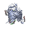 7nrfS S: Starting model for refinement C: citing same article ( |
|---|---|
| Similar structure data |
-Links
 PDBj
PDBj- Assembly
| Deposited unit | 
| ||||||||
|---|---|---|---|---|---|---|---|---|---|
| 1 |
| ||||||||
| Unit cell |
|
-Components
| #1: Protein | Mass: 43805.457 Da / Num. of mol.: 1 Source method: isolated from a genetically manipulated source Source: (gene. exp.) |
|---|---|
| #2: Protein/peptide | Type: Peptide-like / Class: Antibiotic / Mass: 1311.405 Da / Num. of mol.: 1 / Source method: obtained synthetically / Source: (synth.) Photorhabdus australis (bacteria) / References: UniProt: A0A1C0U7H2, BIRD: PRD_002404 |
| #3: Water | ChemComp-HOH /  Mass: 18.015 Da / Num. of mol.: 77 / Source method: isolated from a natural source / Formula: H2O Mass: 18.015 Da / Num. of mol.: 77 / Source method: isolated from a natural source / Formula: H2O |
| Compound details | Dynobactin A is a decapeptide (1-WNSNVHSYRF-10) with 2 closed rings. One carbon-carbon bond formed ...Dynobactin A is a decapeptide (1-WNSNVHSYRF |
| Has ligand of interest | Y |
-Experimental details
-Experiment
| Experiment | Method: X-RAY DIFFRACTION / Number of used crystals: 1 |
|---|
- Sample preparation
| Crystal | Density Matthews: 3.12 Å3/Da / Density % sol: 60.59 % |
|---|---|
| Crystal grow | Temperature: 298 K / Method: vapor diffusion Details: 0.12 M lithium sulfate, 0.02 M Tris pH7.5, 0.1 M sodium citrate pH 5.0, and 20% (v/v) PEG300 |
-Data collection
| Diffraction | Mean temperature: 100 K / Serial crystal experiment: N |
|---|---|
| Diffraction source | Source: SYNCHROTRON / Site: SLS / Beamline: X06DA / Wavelength: 1.000003 Å |
| Detector | Type: DECTRIS PILATUS 2M-F / Detector: PIXEL / Date: Dec 18, 2020 |
| Radiation | Protocol: SINGLE WAVELENGTH / Monochromatic (M) / Laue (L): M / Scattering type: x-ray |
| Radiation wavelength | Wavelength: 1.000003 Å / Relative weight: 1 |
| Reflection | Resolution: 2.5→48.36 Å / Num. obs: 19622 / % possible obs: 99 % / Redundancy: 27.9 % / CC1/2: 0.998 / Rmerge(I) obs: 0.217 / Rpim(I) all: 0.044 / Net I/σ(I): 9 |
| Reflection shell | Resolution: 2.5→2.6 Å / Rmerge(I) obs: 4.571 / Mean I/σ(I) obs: 1 / Num. unique obs: 2171 / CC1/2: 0.718 / Rpim(I) all: 0.893 |
- Processing
| Software |
| |||||||||||||||||||||||||||||||||||||||||||||||||||||||||||||||||||||||||||||||||||||||||||||||||||||||||||||||||||||||||||||
|---|---|---|---|---|---|---|---|---|---|---|---|---|---|---|---|---|---|---|---|---|---|---|---|---|---|---|---|---|---|---|---|---|---|---|---|---|---|---|---|---|---|---|---|---|---|---|---|---|---|---|---|---|---|---|---|---|---|---|---|---|---|---|---|---|---|---|---|---|---|---|---|---|---|---|---|---|---|---|---|---|---|---|---|---|---|---|---|---|---|---|---|---|---|---|---|---|---|---|---|---|---|---|---|---|---|---|---|---|---|---|---|---|---|---|---|---|---|---|---|---|---|---|---|---|---|---|
| Refinement | Method to determine structure: MOLECULAR REPLACEMENT Starting model: 7NRF Resolution: 2.5→48.36 Å / Cor.coef. Fo:Fc: 0.884 / Cor.coef. Fo:Fc free: 0.906 / Cross valid method: THROUGHOUT / σ(F): 0 / SU R Blow DPI: 0.406 / SU Rfree Blow DPI: 0.27 Details: HYDROGENS WERE FULLY REFINED WITH ZERO OCCUPANCY AT NUCLEAR POSITION. REFINEMENT NOTES. NUMBER OF REFINEMENT NOTES : 1 NOTE 1 : IDEAL-DIST CONTACT TERM CONTACT SETUP. ALL ATOMS HAVE CCP4 ATOM TYPE FROM LIBRARY
| |||||||||||||||||||||||||||||||||||||||||||||||||||||||||||||||||||||||||||||||||||||||||||||||||||||||||||||||||||||||||||||
| Displacement parameters | Biso max: 310.76 Å2 / Biso mean: 113.32 Å2 / Biso min: 41.78 Å2
| |||||||||||||||||||||||||||||||||||||||||||||||||||||||||||||||||||||||||||||||||||||||||||||||||||||||||||||||||||||||||||||
| Refine analyze | Luzzati coordinate error obs: 0.51 Å | |||||||||||||||||||||||||||||||||||||||||||||||||||||||||||||||||||||||||||||||||||||||||||||||||||||||||||||||||||||||||||||
| Refinement step | Cycle: final / Resolution: 2.5→48.36 Å
| |||||||||||||||||||||||||||||||||||||||||||||||||||||||||||||||||||||||||||||||||||||||||||||||||||||||||||||||||||||||||||||
| Refine LS restraints |
| |||||||||||||||||||||||||||||||||||||||||||||||||||||||||||||||||||||||||||||||||||||||||||||||||||||||||||||||||||||||||||||
| LS refinement shell | Resolution: 2.5→2.52 Å / Rfactor Rfree error: 0 / Total num. of bins used: 50
| |||||||||||||||||||||||||||||||||||||||||||||||||||||||||||||||||||||||||||||||||||||||||||||||||||||||||||||||||||||||||||||
| Refinement TLS params. | Method: refined / Refine-ID: X-RAY DIFFRACTION
| |||||||||||||||||||||||||||||||||||||||||||||||||||||||||||||||||||||||||||||||||||||||||||||||||||||||||||||||||||||||||||||
| Refinement TLS group |
|
