 Movie
Movie Controller

[English] 日本語
 Yorodumi
- PDB-7r1m: Crystal structure of ExsFA, a Bacillus cereus spore exosporium protein -
+ Open data

- Basic information
| Entry | Database: PDB / ID: 7r1m | ||||||
|---|---|---|---|---|---|---|---|
| Title | Crystal structure of ExsFA, a Bacillus cereus spore exosporium protein | ||||||
 Components Components | Exosporium protein | ||||||
 Keywords Keywords | STRUCTURAL PROTEIN / ExsFA / BxpB / Bacillus cereus / exosporium / endospore | ||||||
| Function / homology | metal ion binding / : / Exosporium protein Function and homology information Function and homology information | ||||||
| Biological species |  | ||||||
| Method |  X-RAY DIFFRACTION / SYNCHROTRON / SAD / molecular replacement / Resolution: 1.64 Å X-RAY DIFFRACTION / SYNCHROTRON / SAD / molecular replacement / Resolution: 1.64 Å | ||||||
 Authors Authors | Brear, P. / Schack, S. / Christie, G. | ||||||
| Funding support |  United Kingdom, 1items United Kingdom, 1items
| ||||||
 Citation Citation | Journal: To Be Published Title: Crystal structure of ExsFA, a Bacillus cereus spore exosporium protein" Authors: Brear, P. / Schack, S. / Christie, G. | ||||||
| History |
|
- Structure visualization
| Structure viewer | Molecule: MolmilJmol/JSmol |
|---|
- Downloads & links
-Download
| PDBx/mmCIF format | 7r1m.cif.gz | 105 KB | Display | PDBx/mmCIF format |
|---|---|---|---|---|
| PDB format | pdb7r1m.ent.gz | 79.6 KB | Display | PDB format |
| PDBx/mmJSON format | 7r1m.json.gz | Tree view | PDBx/mmJSON format | |
| Others |  Other downloads Other downloads |
-Validation report
| Arichive directory | https://data.pdbj.org/pub/pdb/validation_reports/r1/7r1mftp://data.pdbj.org/pub/pdb/validation_reports/r1/7r1m | HTTPS FTP |
|---|
-Related structure data

-Links
 PDBj
PDBj

- Assembly
| Deposited unit | 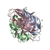
| ||||||||
|---|---|---|---|---|---|---|---|---|---|
| 1 |
| ||||||||
| Unit cell |
|
-Components
-Protein , 1 types, 3 molecules ABC
| #1: Protein | Mass: 18674.137 Da / Num. of mol.: 3 / Mutation: C5S, C12S Source method: isolated from a genetically manipulated source Source: (gene. exp.) Gene: exsF, AT268_09550, COC69_11355, COK98_01700, COM79_21560, D0437_05965, FC702_13950, NCTC7464_04016 Plasmid: pET28a / Production host: |
|---|
-Non-polymers , 5 types, 321 molecules 


| #2: Chemical | ChemComp-AU /  Mass: 196.967 Da / Num. of mol.: 13 / Source method: obtained synthetically / Formula: Au Mass: 196.967 Da / Num. of mol.: 13 / Source method: obtained synthetically / Formula: Au#3: Chemical |  Mass: 24.305 Da / Num. of mol.: 2 / Source method: obtained synthetically / Formula: Mg Mass: 24.305 Da / Num. of mol.: 2 / Source method: obtained synthetically / Formula: Mg#4: Chemical | ChemComp-CA / |  Mass: 40.078 Da / Num. of mol.: 1 / Source method: obtained synthetically / Formula: Ca Mass: 40.078 Da / Num. of mol.: 1 / Source method: obtained synthetically / Formula: Ca#5: Chemical | ChemComp-BTB / |  Mass: 209.240 Da / Num. of mol.: 1 / Source method: obtained synthetically / Formula: C8H19NO5 / Comment: pH buffer*YM Mass: 209.240 Da / Num. of mol.: 1 / Source method: obtained synthetically / Formula: C8H19NO5 / Comment: pH buffer*YM#6: Water | ChemComp-HOH / | Mass: 18.015 Da / Num. of mol.: 304 / Source method: isolated from a natural source / Formula: H2O |
|---|
-Details
| Has ligand of interest | N |
|---|
-Experimental details
-Experiment
| Experiment | Method: X-RAY DIFFRACTION / Number of used crystals: 1 |
|---|
- Sample preparation
| Crystal | Density Matthews: 2.58 Å3/Da / Density % sol: 52.3 % |
|---|---|
| Crystal grow | Temperature: 292 K / Method: vapor diffusion, sitting drop / pH: 7.6 / Details: 22.5% (v/v) PEGSH, 0.2 M KCl |
-Data collection
| Diffraction | Mean temperature: 100 K / Serial crystal experiment: N | ||||||||||||||||||||||||
|---|---|---|---|---|---|---|---|---|---|---|---|---|---|---|---|---|---|---|---|---|---|---|---|---|---|
| Diffraction source | Source: SYNCHROTRON / Site: Diamond / Beamline: I04 / Wavelength: 0.9795 Å | ||||||||||||||||||||||||
| Detector | Type: DECTRIS EIGER2 X 16M / Detector: PIXEL / Date: Oct 9, 2021 | ||||||||||||||||||||||||
| Radiation | Protocol: SINGLE WAVELENGTH / Monochromatic (M) / Laue (L): M / Scattering type: x-ray | ||||||||||||||||||||||||
| Radiation wavelength | Wavelength: 0.9795 Å / Relative weight: 1 | ||||||||||||||||||||||||
| Reflection | Resolution: 1.64→67.39 Å / Num. obs: 66528 / % possible obs: 100 % / Redundancy: 86.7 % / Biso Wilson estimate: 29.51 Å2 / Rpim(I) all: 0.021 / Rrim(I) all: 0.199 / Net I/σ(I): 19.2 / Num. measured all: 5766918 | ||||||||||||||||||||||||
| Reflection shell | Diffraction-ID: 1
|
-Phasing
| Phasing | Method: molecular replacement |
|---|
- Processing
| Software |
| ||||||||||||||||||||||||
|---|---|---|---|---|---|---|---|---|---|---|---|---|---|---|---|---|---|---|---|---|---|---|---|---|---|
| Refinement | Method to determine structure: SAD / Resolution: 1.64→67.39 Å / Cor.coef. Fo:Fc: 0.961 / Cor.coef. Fo:Fc free: 0.952 / SU B: 0.003 / SU ML: 0 / Cross valid method: THROUGHOUT / σ(F): 0 / ESU R: 0.085 / ESU R Free: 0.096 / Stereochemistry target values: MAXIMUM LIKELIHOOD Details: HYDROGENS HAVE BEEN ADDED IN THE RIDING POSITIONS U VALUES : REFINED INDIVIDUALLY
| ||||||||||||||||||||||||
| Solvent computation | Ion probe radii: 0.8 Å / Shrinkage radii: 0.8 Å / VDW probe radii: 1.2 Å / Solvent model: MASK | ||||||||||||||||||||||||
| Displacement parameters | Biso max: 157.65 Å2 / Biso mean: 39.245 Å2 / Biso min: 20.91 Å2
| ||||||||||||||||||||||||
| Refinement step | Cycle: final / Resolution: 1.64→67.39 Å
| ||||||||||||||||||||||||
| LS refinement shell | Resolution: 1.641→1.683 Å / Rfactor Rfree error: 0 / Total num. of bins used: 20
|
