 Movie
Movie Controller


+ Open data

- Basic information
| Entry | Database: PDB / ID: 7r0t | ||||||
|---|---|---|---|---|---|---|---|
| Title | Crystal structure of exonuclease ExnV1 | ||||||
 Components Components | Exonuclease ExnV1 | ||||||
 Keywords Keywords | VIRAL PROTEIN / exonuclease | ||||||
| Function / homology | TERBIUM(III) ION / THYMIDINE-5'-PHOSPHATE Function and homology information Function and homology information | ||||||
| Biological species |   Thermus phage TSP4 (virus) Thermus phage TSP4 (virus) | ||||||
| Method |  X-RAY DIFFRACTION / SYNCHROTRON / SAD / Resolution: 2.194 Å X-RAY DIFFRACTION / SYNCHROTRON / SAD / Resolution: 2.194 Å | ||||||
 Authors Authors | Welin, M. / Svensson, A. / Hakansson, M. / Al-Karadaghi, S. / Jasilionis, A. / Linares-Pasten, J.A. / Wang, L. / Nordberg Karlsson, E. / Ahlqvist, J. | ||||||
| Funding support | European Union, 1items
| ||||||
 Citation Citation | Journal: Acta Crystallogr D Struct Biol / Year: 2022 Title: Crystal structure of DNA polymerase I from Thermus phage G20c. Authors: Ahlqvist, J. / Linares-Pasten, J.A. / Jasilionis, A. / Welin, M. / Hakansson, M. / Svensson, L.A. / Wang, L. / Watzlawick, H. / Aevarsson, A. / Fridjonsson, O.H. / Hreggvidsson, G.O. / ...Authors: Ahlqvist, J. / Linares-Pasten, J.A. / Jasilionis, A. / Welin, M. / Hakansson, M. / Svensson, L.A. / Wang, L. / Watzlawick, H. / Aevarsson, A. / Fridjonsson, O.H. / Hreggvidsson, G.O. / Ketelsen Striberny, B. / Glomsaker, E. / Lanes, O. / Al-Karadaghi, S. / Nordberg Karlsson, E. | ||||||
| History |
|
- Structure visualization
| Structure viewer | Molecule: MolmilJmol/JSmol |
|---|
- Downloads & links
-Download
| PDBx/mmCIF format | 7r0t.cif.gz | 132.7 KB | Display | PDBx/mmCIF format |
|---|---|---|---|---|
| PDB format | pdb7r0t.ent.gz | 103.4 KB | Display | PDB format |
| PDBx/mmJSON format | 7r0t.json.gz | Tree view | PDBx/mmJSON format | |
| Others |  Other downloads Other downloads |
-Validation report
| Summary document | 7r0t_validation.pdf.gz | 831.7 KB | Display | wwPDB validaton report |
|---|---|---|---|---|
| Full document | 7r0t_full_validation.pdf.gz | 834 KB | Display | |
| Data in XML | 7r0t_validation.xml.gz | 13 KB | Display | |
| Data in CIF | 7r0t_validation.cif.gz | 17.4 KB | Display | |
| Arichive directory | https://data.pdbj.org/pub/pdb/validation_reports/r0/7r0tftp://data.pdbj.org/pub/pdb/validation_reports/r0/7r0t | HTTPS FTP |
-Related structure data

-Links
 PDBj
PDBj- Assembly
| Deposited unit | 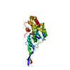
| ||||||||
|---|---|---|---|---|---|---|---|---|---|
| 1 |
| ||||||||
| Unit cell |
|
-Components
-Protein , 1 types, 1 molecules A
| #1: Protein | Mass: 36785.906 Da / Num. of mol.: 1 Source method: isolated from a genetically manipulated source Source: (gene. exp.) Thermus phage TSP4 (virus) / Production host:  |
|---|
-Non-polymers , 5 types, 54 molecules 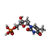


| #2: Chemical | ChemComp-TMP /  Mass: 322.208 Da / Num. of mol.: 1 / Source method: obtained synthetically / Formula: C10H15N2O8P / Feature type: SUBJECT OF INVESTIGATION Mass: 322.208 Da / Num. of mol.: 1 / Source method: obtained synthetically / Formula: C10H15N2O8P / Feature type: SUBJECT OF INVESTIGATION | ||||||
|---|---|---|---|---|---|---|---|
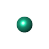
| #3: Chemical |  Mass: 24.305 Da / Num. of mol.: 2 / Source method: obtained synthetically / Formula: Mg Mass: 24.305 Da / Num. of mol.: 2 / Source method: obtained synthetically / Formula: Mg#4: Chemical | ChemComp-TB / |  Mass: 158.925 Da / Num. of mol.: 1 / Source method: obtained synthetically / Formula: Tb Mass: 158.925 Da / Num. of mol.: 1 / Source method: obtained synthetically / Formula: Tb#5: Chemical | ChemComp-CL /  Mass: 35.453 Da / Num. of mol.: 4 / Source method: obtained synthetically / Formula: Cl Mass: 35.453 Da / Num. of mol.: 4 / Source method: obtained synthetically / Formula: Cl#6: Water | ChemComp-HOH / | Mass: 18.015 Da / Num. of mol.: 46 / Source method: isolated from a natural source / Formula: H2O |
-Details
| Has ligand of interest | Y |
|---|
-Experimental details
-Experiment
| Experiment | Method: X-RAY DIFFRACTION / Number of used crystals: 1 |
|---|
- Sample preparation
| Crystal | Density Matthews: 2.67 Å3/Da / Density % sol: 54 % |
|---|---|
| Crystal grow | Temperature: 277 K / Method: vapor diffusion, sitting drop / pH: 9.3 / Details: 0.1 M Bicine pH 9.3 30 % PEG Smear Low |
-Data collection
| Diffraction | Mean temperature: 100 K / Serial crystal experiment: N |
|---|---|
| Diffraction source | Source: SYNCHROTRON / Site: Diamond  / Beamline: I03 / Wavelength: 1.6488 Å / Beamline: I03 / Wavelength: 1.6488 Å |
| Detector | Type: DECTRIS EIGER2 XE 16M / Detector: PIXEL / Date: Feb 15, 2019 |
| Radiation | Protocol: SINGLE WAVELENGTH / Monochromatic (M) / Laue (L): M / Scattering type: x-ray |
| Radiation wavelength | Wavelength: 1.6488 Å / Relative weight: 1 |
| Reflection | Resolution: 2.19→45.78 Å / Num. obs: 20566 / % possible obs: 98.1 % / Redundancy: 12.7 % / CC1/2: 0.999 / Rmerge(I) obs: 0.136 / Rpim(I) all: 0.04 / Rrim(I) all: 0.141 / Net I/σ(I): 12.5 |
| Reflection shell | Resolution: 2.19→2.26 Å / Rmerge(I) obs: 3.187 / Mean I/σ(I) obs: 1.1 / Num. unique obs: 1650 / CC1/2: 0.792 / Rpim(I) all: 0.935 / Rrim(I) all: 3.325 |
- Processing
| Software |
| ||||||||||||||||||||||||||||||||||||||||||||||||||||||||||||||||||
|---|---|---|---|---|---|---|---|---|---|---|---|---|---|---|---|---|---|---|---|---|---|---|---|---|---|---|---|---|---|---|---|---|---|---|---|---|---|---|---|---|---|---|---|---|---|---|---|---|---|---|---|---|---|---|---|---|---|---|---|---|---|---|---|---|---|---|---|
| Refinement | Method to determine structure: SAD / Resolution: 2.194→45.78 Å / Cor.coef. Fo:Fc: 0.935 / Cor.coef. Fo:Fc free: 0.918 / SU R Cruickshank DPI: 0.244 / Cross valid method: THROUGHOUT / SU R Blow DPI: 0.244 / SU Rfree Blow DPI: 0.209 / SU Rfree Cruickshank DPI: 0.21
| ||||||||||||||||||||||||||||||||||||||||||||||||||||||||||||||||||
| Displacement parameters | Biso mean: 62.27 Å2
| ||||||||||||||||||||||||||||||||||||||||||||||||||||||||||||||||||
| Refine analyze | Luzzati coordinate error obs: 0.38 Å | ||||||||||||||||||||||||||||||||||||||||||||||||||||||||||||||||||
| Refinement step | Cycle: LAST / Resolution: 2.194→45.78 Å
| ||||||||||||||||||||||||||||||||||||||||||||||||||||||||||||||||||
| Refine LS restraints |
| ||||||||||||||||||||||||||||||||||||||||||||||||||||||||||||||||||
| LS refinement shell | Resolution: 2.194→2.21 Å
| ||||||||||||||||||||||||||||||||||||||||||||||||||||||||||||||||||
| Refinement TLS params. | Origin x: 20.2496 Å / Origin y: 16.6061 Å / Origin z: 142.517 Å
| ||||||||||||||||||||||||||||||||||||||||||||||||||||||||||||||||||
| Refinement TLS group | Selection details: { A|0 - 296 } |
