Movie
Movie Controller


+ Open data

- Basic information
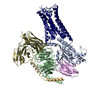
| Entry | Database: PDB / ID: 7r0c | |||||||||||||||||||||||||||||||||||||||||||||||||||
|---|---|---|---|---|---|---|---|---|---|---|---|---|---|---|---|---|---|---|---|---|---|---|---|---|---|---|---|---|---|---|---|---|---|---|---|---|---|---|---|---|---|---|---|---|---|---|---|---|---|---|---|---|
| Title | Structure of the AVP-V2R-arrestin2-ScFv30 complex | |||||||||||||||||||||||||||||||||||||||||||||||||||
 Components Components |
| |||||||||||||||||||||||||||||||||||||||||||||||||||
 Keywords Keywords | MEMBRANE PROTEIN / G-protein coupled receptor V2 receptor Arrestin 2 Vasopressin | |||||||||||||||||||||||||||||||||||||||||||||||||||
| Function / homology |  Function and homology information Function and homology informationrenal water retention / Defective AVP does not bind AVPR2 and causes neurohypophyseal diabetes insipidus (NDI) / regulation of systemic arterial blood pressure by vasopressin / Vasopressin-like receptors / vasopressin receptor activity / renal water absorption / angiotensin receptor binding / TGFBR3 regulates TGF-beta signaling / Activation of SMO / hemostasis ...renal water retention / Defective AVP does not bind AVPR2 and causes neurohypophyseal diabetes insipidus (NDI) / regulation of systemic arterial blood pressure by vasopressin / Vasopressin-like receptors / vasopressin receptor activity / renal water absorption / angiotensin receptor binding / TGFBR3 regulates TGF-beta signaling / Activation of SMO / hemostasis / telencephalon development / negative regulation of interleukin-8 production / desensitization of G protein-coupled receptor signaling pathway / arrestin family protein binding / G protein-coupled receptor internalization / : / sensory perception / Lysosome Vesicle Biogenesis / positive regulation of cardiac muscle hypertrophy / stress fiber assembly / Golgi Associated Vesicle Biogenesis / positive regulation of Rho protein signal transduction / positive regulation of systemic arterial blood pressure / negative regulation of interleukin-6 production / pseudopodium / positive regulation of intracellular signal transduction / negative regulation of Notch signaling pathway / positive regulation of receptor internalization / positive regulation of vasoconstriction / endocytic vesicle / activation of adenylate cyclase activity / cellular response to hormone stimulus / insulin-like growth factor receptor binding / clathrin-coated pit / response to cytokine / negative regulation of protein ubiquitination / intracellular glucose homeostasis / cytoplasmic vesicle membrane / enzyme inhibitor activity / Activated NOTCH1 Transmits Signal to the Nucleus / GTPase activator activity / clathrin-coated endocytic vesicle membrane / Signaling by high-kinase activity BRAF mutants / MAP2K and MAPK activation / positive regulation of protein phosphorylation / G protein-coupled receptor binding / adenylate cyclase-modulating G protein-coupled receptor signaling pathway / Signaling by RAF1 mutants / Signaling by moderate kinase activity BRAF mutants / Paradoxical activation of RAF signaling by kinase inactive BRAF / Signaling downstream of RAS mutants / endocytic vesicle membrane / Signaling by BRAF and RAF1 fusions / Vasopressin regulates renal water homeostasis via Aquaporins / Cargo recognition for clathrin-mediated endocytosis / protein transport / Clathrin-mediated endocytosis / Thrombin signalling through proteinase activated receptors (PARs) / adenylate cyclase-activating G protein-coupled receptor signaling pathway / cytoplasmic vesicle / molecular adaptor activity / G alpha (s) signalling events / ubiquitin-dependent protein catabolic process / proteasome-mediated ubiquitin-dependent protein catabolic process / positive regulation of ERK1 and ERK2 cascade / transcription coactivator activity / cell surface receptor signaling pathway / endosome / nuclear body / Ub-specific processing proteases / protein ubiquitination / G protein-coupled receptor signaling pathway / negative regulation of cell population proliferation / Golgi membrane / lysosomal membrane / positive regulation of cell population proliferation / positive regulation of gene expression / ubiquitin protein ligase binding / regulation of transcription by RNA polymerase II / chromatin / perinuclear region of cytoplasm / Golgi apparatus / endoplasmic reticulum / positive regulation of transcription by RNA polymerase II / nucleoplasm / membrane / nucleus / plasma membrane / cytosol / cytoplasm Similarity search - Function | |||||||||||||||||||||||||||||||||||||||||||||||||||
| Biological species |  Homo sapiens (human) Homo sapiens (human)synthetic construct (others) | |||||||||||||||||||||||||||||||||||||||||||||||||||
| Method | ELECTRON MICROSCOPY / single particle reconstruction / cryo EM / Resolution: 4.73 Å | |||||||||||||||||||||||||||||||||||||||||||||||||||
 Authors Authors | Bous, J. / Fouillen, A. / Trapani, S. / Granier, S. / Mouillac, B. / Bron, P. | |||||||||||||||||||||||||||||||||||||||||||||||||||
| Funding support |  France, 2items France, 2items
| |||||||||||||||||||||||||||||||||||||||||||||||||||
 Citation Citation | Journal: Sci Adv / Year: 2022 Title: Structure of the vasopressin hormone-V2 receptor-β-arrestin1 ternary complex. Authors: Julien Bous / Aurélien Fouillen / Hélène Orcel / Stefano Trapani / Xiaojing Cong / Simon Fontanel / Julie Saint-Paul / Joséphine Lai-Kee-Him / Serge Urbach / Nathalie Sibille / Rémy ...Authors: Julien Bous / Aurélien Fouillen / Hélène Orcel / Stefano Trapani / Xiaojing Cong / Simon Fontanel / Julie Saint-Paul / Joséphine Lai-Kee-Him / Serge Urbach / Nathalie Sibille / Rémy Sounier / Sébastien Granier / Bernard Mouillac / Patrick Bron / Abstract: Arrestins interact with G protein-coupled receptors (GPCRs) to stop G protein activation and to initiate key signaling pathways. Recent structural studies shed light on the molecular mechanisms ...Arrestins interact with G protein-coupled receptors (GPCRs) to stop G protein activation and to initiate key signaling pathways. Recent structural studies shed light on the molecular mechanisms involved in GPCR-arrestin coupling, but whether this process is conserved among GPCRs is poorly understood. Here, we report the cryo-electron microscopy active structure of the wild-type arginine-vasopressin V2 receptor (V2R) in complex with β-arrestin1. It reveals an atypical position of β-arrestin1 compared to previously described GPCR-arrestin assemblies, associated with an original V2R/β-arrestin1 interface involving all receptor intracellular loops. Phosphorylated sites of the V2R carboxyl terminus are clearly identified and interact extensively with the β-arrestin1 N-lobe, in agreement with structural data obtained with chimeric or synthetic systems. Overall, these findings highlight a notable structural variability among GPCR-arrestin signaling complexes. | |||||||||||||||||||||||||||||||||||||||||||||||||||
| History |
|
- Structure visualization
| Structure viewer | Molecule: MolmilJmol/JSmol |
|---|
- Downloads & links
-Download
| PDBx/mmCIF format | 7r0c.cif.gz | 171.6 KB | Display | PDBx/mmCIF format |
|---|---|---|---|---|
| PDB format | pdb7r0c.ent.gz | 129.6 KB | Display | PDB format |
| PDBx/mmJSON format | 7r0c.json.gz | Tree view | PDBx/mmJSON format | |
| Others |  Other downloads Other downloads |
-Validation report
| Arichive directory | https://data.pdbj.org/pub/pdb/validation_reports/r0/7r0cftp://data.pdbj.org/pub/pdb/validation_reports/r0/7r0c | HTTPS FTP |
|---|
-Related structure data
| Related structure data |  14221MC  7r0jC M: map data used to model this data C: citing same article ( |
|---|---|
| Similar structure data |

-Links
 PDBj
PDBj


- Assembly
| Deposited unit | 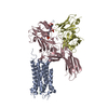
|
|---|---|
| 1 |
|
-Components
| #1: Protein | Mass: 41532.367 Da / Num. of mol.: 1 Source method: isolated from a genetically manipulated source Source: (gene. exp.) Homo sapiens (human) / Gene: AVPR2, ADHR, DIR, DIR3, V2RProduction host: Insect expression vector pBlueBacmsGCA1 (others) References: UniProt: P30518 |
|---|---|
| #2: Protein/peptide | Mass: 1086.248 Da / Num. of mol.: 1 / Source method: obtained synthetically / Source: (synth.) Homo sapiens (human) |
| #3: Protein | Mass: 47164.609 Da / Num. of mol.: 1 Source method: isolated from a genetically manipulated source Source: (gene. exp.) Homo sapiens (human) / Production host:  |
| #4: Antibody | Mass: 30070.027 Da / Num. of mol.: 1 Source method: isolated from a genetically manipulated source Source: (gene. exp.) synthetic construct (others) Production host: Insect expression vector pBlueBacmsGCA1 (others) |
| Has ligand of interest | N |
| Has protein modification | Y |
-Experimental details
-Experiment
| Experiment | Method: ELECTRON MICROSCOPY |
|---|---|
| EM experiment | Aggregation state: PARTICLE / 3D reconstruction method: single particle reconstruction |
- Sample preparation
| Component | Name: Ternary complex of the AVP-V2 receptor with arrestin2 and ScfV30 Type: COMPLEX / Entity ID: all / Source: MULTIPLE SOURCES |
|---|---|
| Molecular weight | Experimental value: NO |
| Source (natural) | Organism: Homo sapiens (human) |
| Source (recombinant) | Organism: Insect expression vector pBlueBacmsGCA1s |
| Buffer solution | pH: 7.5 |
| Specimen | Conc.: 3 mg/ml / Embedding applied: NO / Shadowing applied: NO / Staining applied: NO / Vitrification applied: YES |
| Specimen support | Grid material: GOLD / Grid mesh size: 300 divisions/in. / Grid type: UltrAuFoil R1.2/1.3 |
| Vitrification | Instrument: LEICA EM GP / Cryogen name: ETHANE / Humidity: 100 % / Chamber temperature: 277 K |
- Electron microscopy imaging
| Experimental equipment |  Model: Titan Krios / Image courtesy: FEI Company |
|---|---|
| Microscopy | Model: FEI TITAN KRIOS |
| Electron gun | Electron source:  FIELD EMISSION GUN / Accelerating voltage: 300 kV / Illumination mode: FLOOD BEAM FIELD EMISSION GUN / Accelerating voltage: 300 kV / Illumination mode: FLOOD BEAM |
| Electron lens | Mode: BRIGHT FIELD / Calibrated magnification: 130000 X / Nominal defocus max: 2000 nm / Nominal defocus min: 1000 nm / Cs: 2.7 mm / C2 aperture diameter: 50 µm |
| Specimen holder | Cryogen: NITROGEN |
| Image recording | Average exposure time: 1 sec. / Electron dose: 52 e/Å2 / Film or detector model: GATAN K3 BIOQUANTUM (6k x 4k) / Num. of real images: 14080 |
- Processing
| Software | Name: PHENIX / Version: 1.20_4459: / Classification: refinement | ||||||||||||||||||||||||
|---|---|---|---|---|---|---|---|---|---|---|---|---|---|---|---|---|---|---|---|---|---|---|---|---|---|
| EM software |
| ||||||||||||||||||||||||
| CTF correction | Type: PHASE FLIPPING AND AMPLITUDE CORRECTION | ||||||||||||||||||||||||
| Particle selection | Num. of particles selected: 4595394 | ||||||||||||||||||||||||
| 3D reconstruction | Resolution: 4.73 Å / Resolution method: FSC 0.143 CUT-OFF / Num. of particles: 8296 / Symmetry type: POINT | ||||||||||||||||||||||||
| Atomic model building | 3D fitting-ID: 1 / Source name: PDB / Type: experimental model
| ||||||||||||||||||||||||
| Refine LS restraints |
|