 Movie
Movie Controller

[English] 日本語
 Yorodumi
- PDB-7r09: Amine Dehydrogenase MATOUAmDH2 in complex with NADP+ and Cyclohex... -
+ Open data

- Basic information
| Entry | Database: PDB / ID: 7r09 | ||||||
|---|---|---|---|---|---|---|---|
| Title | Amine Dehydrogenase MATOUAmDH2 in complex with NADP+ and Cyclohexylamine | ||||||
 Components Components | Amine Dehydrogenase | ||||||
 Keywords Keywords | OXIDOREDUCTASE / Amine / NADP | ||||||
| Function / homology | CYCLOHEXYLAMMONIUM ION / NADP NICOTINAMIDE-ADENINE-DINUCLEOTIDE PHOSPHATE Function and homology information Function and homology information | ||||||
| Biological species | metagenome (others) | ||||||
| Method |  X-RAY DIFFRACTION / SYNCHROTRON / MOLECULAR REPLACEMENT / Resolution: 2.08 Å X-RAY DIFFRACTION / SYNCHROTRON / MOLECULAR REPLACEMENT / Resolution: 2.08 Å | ||||||
 Authors Authors | Bennett, M. / Ducrot, L. / Vaxelaire-Vergne, C. / Grogan, G. | ||||||
| Funding support | 1items
| ||||||
 Citation Citation | Journal: Chembiochem / Year: 2022 Title: Structure and Mutation of the Native Amine Dehydrogenase MATOUAmDH2. Authors: Bennett, M. / Ducrot, L. / Vergne-Vaxelaire, C. / Grogan, G. | ||||||
| History |
|
- Structure visualization
| Structure viewer | Molecule: MolmilJmol/JSmol |
|---|
- Downloads & links
-Download
| PDBx/mmCIF format | 7r09.cif.gz | 81.9 KB | Display | PDBx/mmCIF format |
|---|---|---|---|---|
| PDB format | pdb7r09.ent.gz | 59.6 KB | Display | PDB format |
| PDBx/mmJSON format | 7r09.json.gz | Tree view | PDBx/mmJSON format | |
| Others |  Other downloads Other downloads |
-Validation report
| Summary document | 7r09_validation.pdf.gz | 1 MB | Display | wwPDB validaton report |
|---|---|---|---|---|
| Full document | 7r09_full_validation.pdf.gz | 1 MB | Display | |
| Data in XML | 7r09_validation.xml.gz | 15.2 KB | Display | |
| Data in CIF | 7r09_validation.cif.gz | 20.8 KB | Display | |
| Arichive directory | https://data.pdbj.org/pub/pdb/validation_reports/r0/7r09ftp://data.pdbj.org/pub/pdb/validation_reports/r0/7r09 | HTTPS FTP |
-Related structure data
| Related structure data | 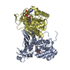 7zboC 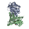 6iauS S: Starting model for refinement C: citing same article ( |
|---|---|
| Similar structure data |
-Links
 PDBj
PDBj- Assembly
| Deposited unit | 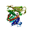
| ||||||||
|---|---|---|---|---|---|---|---|---|---|
| 1 | 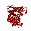
| ||||||||
| Unit cell |
|
-Components
| #1: Protein | Mass: 38249.332 Da / Num. of mol.: 1 Source method: isolated from a genetically manipulated source Source: (gene. exp.) metagenome (others) / Production host:  |
|---|---|
| #2: Chemical | ChemComp-NAP /   Mass: 743.405 Da / Num. of mol.: 1 / Source method: obtained synthetically / Formula: C21H28N7O17P3 Mass: 743.405 Da / Num. of mol.: 1 / Source method: obtained synthetically / Formula: C21H28N7O17P3 |
| #3: Chemical | ChemComp-HAI /   Mass: 100.182 Da / Num. of mol.: 1 / Source method: obtained synthetically / Formula: C6H14N / Feature type: SUBJECT OF INVESTIGATION Mass: 100.182 Da / Num. of mol.: 1 / Source method: obtained synthetically / Formula: C6H14N / Feature type: SUBJECT OF INVESTIGATION |
| #4: Water | ChemComp-HOH /  Mass: 18.015 Da / Num. of mol.: 81 / Source method: isolated from a natural source / Formula: H2O Mass: 18.015 Da / Num. of mol.: 81 / Source method: isolated from a natural source / Formula: H2O |
| Has ligand of interest | Y |
-Experimental details
-Experiment
| Experiment | Method: X-RAY DIFFRACTION / Number of used crystals: 1 |
|---|
- Sample preparation
| Crystal | Density Matthews: 2.5 Å3/Da / Density % sol: 50.81 % |
|---|---|
| Crystal grow | Temperature: 298 K / Method: vapor diffusion, sitting drop / pH: 6.5 Details: 0.1 M bis-Tris pH 6.5, 25% (w/v) PEG 3350 0.2 M MgCl2, 10 mM NADP, 10 mM n-pentylamine |
-Data collection
| Diffraction | Mean temperature: 120 K / Serial crystal experiment: N |
|---|---|
| Diffraction source | Source: SYNCHROTRON / Site: Diamond  / Beamline: I03 / Wavelength: 0.97625 Å / Beamline: I03 / Wavelength: 0.97625 Å |
| Detector | Type: DECTRIS EIGER2 XE 16M / Detector: PIXEL / Date: Dec 10, 2021 |
| Radiation | Protocol: SINGLE WAVELENGTH / Monochromatic (M) / Laue (L): M / Scattering type: x-ray |
| Radiation wavelength | Wavelength: 0.97625 Å / Relative weight: 1 |
| Reflection | Resolution: 2.08→55.4 Å / Num. obs: 23365 / % possible obs: 99.9 % / Redundancy: 20.2 % / Biso Wilson estimate: 48 Å2 / CC1/2: 1 / Rmerge(I) obs: 0.07 / Rpim(I) all: 0.02 / Net I/σ(I): 24.4 |
| Reflection shell | Resolution: 2.08→2.13 Å / Redundancy: 20.7 % / Rmerge(I) obs: 1.28 / Mean I/σ(I) obs: 2.7 / Num. unique obs: 1768 / CC1/2: 0.94 / Rpim(I) all: 0.41 |
- Processing
| Software |
| ||||||||||||||||||||||||||||||||||||||||||||||||||||||||||||
|---|---|---|---|---|---|---|---|---|---|---|---|---|---|---|---|---|---|---|---|---|---|---|---|---|---|---|---|---|---|---|---|---|---|---|---|---|---|---|---|---|---|---|---|---|---|---|---|---|---|---|---|---|---|---|---|---|---|---|---|---|---|
| Refinement | Method to determine structure: MOLECULAR REPLACEMENT Starting model: 6IAU Resolution: 2.08→55.4 Å / Cor.coef. Fo:Fc: 0.953 / Cor.coef. Fo:Fc free: 0.931 / SU B: 8.784 / SU ML: 0.221 / Cross valid method: THROUGHOUT / σ(F): 0 / ESU R: 0.238 / ESU R Free: 0.219 / Stereochemistry target values: MAXIMUM LIKELIHOOD Details: HYDROGENS HAVE BEEN ADDED IN THE RIDING POSITIONS U VALUES : REFINED INDIVIDUALLY
| ||||||||||||||||||||||||||||||||||||||||||||||||||||||||||||
| Solvent computation | Ion probe radii: 0.8 Å / Shrinkage radii: 0.8 Å / VDW probe radii: 1.2 Å / Solvent model: MASK | ||||||||||||||||||||||||||||||||||||||||||||||||||||||||||||
| Displacement parameters | Biso max: 105.88 Å2 / Biso mean: 54.816 Å2 / Biso min: 28.35 Å2
| ||||||||||||||||||||||||||||||||||||||||||||||||||||||||||||
| Refinement step | Cycle: final / Resolution: 2.08→55.4 Å
| ||||||||||||||||||||||||||||||||||||||||||||||||||||||||||||
| Refine LS restraints |
| ||||||||||||||||||||||||||||||||||||||||||||||||||||||||||||
| LS refinement shell | Resolution: 2.08→2.134 Å / Rfactor Rfree error: 0 / Total num. of bins used: 20
|
