 Movie
Movie Controller


+ Open data

- Basic information
| Entry | Database: PDB / ID: 7qzk | ||||||
|---|---|---|---|---|---|---|---|
| Title | Crystal structure of mouse CNPase catalytic domain, V318I mutant | ||||||
 Components Components | 2',3'-cyclic-nucleotide 3'-phosphodiesterase | ||||||
 Keywords Keywords | HYDROLASE / CNPase / 2H phosphodiesterase / mutation | ||||||
| Function / homology |  Function and homology information Function and homology informationcyclic nucleotide catabolic process / 2',3'-cyclic-nucleotide 3'-phosphodiesterase / 2',3'-cyclic-nucleotide 3'-phosphodiesterase activity / oligodendrocyte differentiation / axonogenesis / adult locomotory behavior / response to toxic substance / melanosome / myelin sheath / extracellular space ...cyclic nucleotide catabolic process / 2',3'-cyclic-nucleotide 3'-phosphodiesterase / 2',3'-cyclic-nucleotide 3'-phosphodiesterase activity / oligodendrocyte differentiation / axonogenesis / adult locomotory behavior / response to toxic substance / melanosome / myelin sheath / extracellular space / RNA binding / membrane / cytoplasm Similarity search - Function | ||||||
| Biological species |  | ||||||
| Method |  X-RAY DIFFRACTION / SYNCHROTRON / MOLECULAR REPLACEMENT / Resolution: 1.66 Å X-RAY DIFFRACTION / SYNCHROTRON / MOLECULAR REPLACEMENT / Resolution: 1.66 Å | ||||||
 Authors Authors | Markusson, S. / Kursula, P. | ||||||
| Funding support | 1items
| ||||||
 Citation Citation | Journal: To Be Published Title: Crystal structure of mouse CNPase catalytic domain, V318I mutant Authors: Markusson, S. / Kursula, P. | ||||||
| History |
|
- Structure visualization
| Structure viewer | Molecule: MolmilJmol/JSmol |
|---|
- Downloads & links
-Download
| PDBx/mmCIF format | 7qzk.cif.gz | 116.3 KB | Display | PDBx/mmCIF format |
|---|---|---|---|---|
| PDB format | pdb7qzk.ent.gz | 72.5 KB | Display | PDB format |
| PDBx/mmJSON format | 7qzk.json.gz | Tree view | PDBx/mmJSON format | |
| Others |  Other downloads Other downloads |
-Validation report
| Summary document | 7qzk_validation.pdf.gz | 441.3 KB | Display | wwPDB validaton report |
|---|---|---|---|---|
| Full document | 7qzk_full_validation.pdf.gz | 443.6 KB | Display | |
| Data in XML | 7qzk_validation.xml.gz | 12 KB | Display | |
| Data in CIF | 7qzk_validation.cif.gz | 16.8 KB | Display | |
| Arichive directory | https://data.pdbj.org/pub/pdb/validation_reports/qz/7qzkftp://data.pdbj.org/pub/pdb/validation_reports/qz/7qzk | HTTPS FTP |
-Related structure data
| Related structure data | 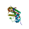 2xmiS S: Starting model for refinement |
|---|---|
| Similar structure data | |
| Experimental dataset #1 | Data reference: 10.5281/zenodo.5323918 / Data set type: diffraction image data |
-Links
 PDBj
PDBj- Assembly
| Deposited unit | 
| ||||||||||||
|---|---|---|---|---|---|---|---|---|---|---|---|---|---|
| 1 |
| ||||||||||||
| Unit cell |
|
-Components
| #1: Protein | Mass: 24307.955 Da / Num. of mol.: 1 Source method: isolated from a genetically manipulated source Source: (gene. exp.)  References: UniProt: P16330, 2',3'-cyclic-nucleotide 3'-phosphodiesterase |
|---|---|
| #2: Chemical | ChemComp-CIT /   Mass: 192.124 Da / Num. of mol.: 1 / Source method: obtained synthetically / Formula: C6H8O7 Mass: 192.124 Da / Num. of mol.: 1 / Source method: obtained synthetically / Formula: C6H8O7 |
| #3: Water | ChemComp-HOH /  Mass: 18.015 Da / Num. of mol.: 152 / Source method: isolated from a natural source / Formula: H2O Mass: 18.015 Da / Num. of mol.: 152 / Source method: isolated from a natural source / Formula: H2O |
| Has ligand of interest | N |
-Experimental details
-Experiment
| Experiment | Method: X-RAY DIFFRACTION / Number of used crystals: 1 |
|---|
- Sample preparation
| Crystal | Density Matthews: 1.97 Å3/Da / Density % sol: 37.71 % |
|---|---|
| Crystal grow | Temperature: 293 K / Method: vapor diffusion, sitting drop / pH: 3.5 / Details: 23% PEG3350 and 50 mM tri-sodium citrate (pH 3.5) |
-Data collection
| Diffraction | Mean temperature: 100 K / Serial crystal experiment: N |
|---|---|
| Diffraction source | Source: SYNCHROTRON / Site: ESRF  / Beamline: MASSIF-3 / Wavelength: 0.968 Å / Beamline: MASSIF-3 / Wavelength: 0.968 Å |
| Detector | Type: DECTRIS EIGER X 4M / Detector: PIXEL / Date: Feb 20, 2021 |
| Radiation | Protocol: SINGLE WAVELENGTH / Monochromatic (M) / Laue (L): M / Scattering type: x-ray |
| Radiation wavelength | Wavelength: 0.968 Å / Relative weight: 1 |
| Reflection | Resolution: 1.66→40 Å / Num. obs: 22318 / % possible obs: 98.9 % / Redundancy: 6.8 % / Biso Wilson estimate: 22.23 Å2 / CC1/2: 0.995 / Rrim(I) all: 0.192 / Net I/σ(I): 6.7 |
| Reflection shell | Resolution: 1.66→1.7 Å / Redundancy: 7 % / Mean I/σ(I) obs: 0.7 / Num. unique obs: 1662 / CC1/2: 0.342 / Rrim(I) all: 2.164 / % possible all: 99.8 |
- Processing
| Software |
| |||||||||||||||||||||||||||||||||||||||||||||||||||||||||||||||||||||||||||||||||||||||||||||||||||||||||
|---|---|---|---|---|---|---|---|---|---|---|---|---|---|---|---|---|---|---|---|---|---|---|---|---|---|---|---|---|---|---|---|---|---|---|---|---|---|---|---|---|---|---|---|---|---|---|---|---|---|---|---|---|---|---|---|---|---|---|---|---|---|---|---|---|---|---|---|---|---|---|---|---|---|---|---|---|---|---|---|---|---|---|---|---|---|---|---|---|---|---|---|---|---|---|---|---|---|---|---|---|---|---|---|---|---|---|
| Refinement | Method to determine structure: MOLECULAR REPLACEMENT Starting model: 2XMI Resolution: 1.66→39.22 Å / SU ML: 0.2612 / Cross valid method: FREE R-VALUE / σ(F): 1.35 / Phase error: 23.0087 Stereochemistry target values: GeoStd + Monomer Library + CDL v1.2
| |||||||||||||||||||||||||||||||||||||||||||||||||||||||||||||||||||||||||||||||||||||||||||||||||||||||||
| Solvent computation | Shrinkage radii: 0.9 Å / VDW probe radii: 1.1 Å / Solvent model: FLAT BULK SOLVENT MODEL | |||||||||||||||||||||||||||||||||||||||||||||||||||||||||||||||||||||||||||||||||||||||||||||||||||||||||
| Displacement parameters | Biso mean: 29.04 Å2 | |||||||||||||||||||||||||||||||||||||||||||||||||||||||||||||||||||||||||||||||||||||||||||||||||||||||||
| Refinement step | Cycle: LAST / Resolution: 1.66→39.22 Å
| |||||||||||||||||||||||||||||||||||||||||||||||||||||||||||||||||||||||||||||||||||||||||||||||||||||||||
| Refine LS restraints |
| |||||||||||||||||||||||||||||||||||||||||||||||||||||||||||||||||||||||||||||||||||||||||||||||||||||||||
| LS refinement shell |
|
