 Movie
Movie Controller

[English] 日本語
 Yorodumi
- PDB-7qze: SFX structure of dye-type peroxidase DtpB D152A variant in the fe... -
+ Open data

- Basic information
| Entry | Database: PDB / ID: 7qze | ||||||
|---|---|---|---|---|---|---|---|
| Title | SFX structure of dye-type peroxidase DtpB D152A variant in the ferryl state | ||||||
 Components Components | Dyp-type peroxidase family | ||||||
 Keywords Keywords | OXIDOREDUCTASE / haem / peroxidase / ferric | ||||||
| Function / homology |  Function and homology information Function and homology information | ||||||
| Biological species |  Streptomyces lividans (bacteria) Streptomyces lividans (bacteria) | ||||||
| Method |  X-RAY DIFFRACTION / FREE ELECTRON LASER / MOLECULAR REPLACEMENT / Resolution: 1.9 Å X-RAY DIFFRACTION / FREE ELECTRON LASER / MOLECULAR REPLACEMENT / Resolution: 1.9 Å | ||||||
 Authors Authors | Lucic, M. / Worrall, J.A.R. / Hough, M.A. / Shilova, A. / Axford, D.A. / Owen, R.L. / Tosha, T. / Sugimoto, H. / Owada, S. | ||||||
| Funding support |  United Kingdom, 1items United Kingdom, 1items
| ||||||
 Citation Citation | Journal: Acs Catalysis / Year: 2022 Title: Serial Femtosecond Crystallography Reveals the Role of Water in the One- or Two-Electron Redox Chemistry of Compound I in the Catalytic Cycle of the B-Type Dye-Decolorizing Peroxidase DtpB. Authors: Lucic, M. / Wilson, M.T. / Tosha, T. / Sugimoto, H. / Shilova, A. / Axford, D. / Owen, R.L. / Hough, M.A. / Worrall, J.A.R. | ||||||
| History |
|
- Structure visualization
| Structure viewer | Molecule: MolmilJmol/JSmol |
|---|
- Downloads & links
-Download
| PDBx/mmCIF format | 7qze.cif.gz | 373.9 KB | Display | PDBx/mmCIF format |
|---|---|---|---|---|
| PDB format | pdb7qze.ent.gz | 303.8 KB | Display | PDB format |
| PDBx/mmJSON format | 7qze.json.gz | Tree view | PDBx/mmJSON format | |
| Others |  Other downloads Other downloads |
-Validation report
| Arichive directory | https://data.pdbj.org/pub/pdb/validation_reports/qz/7qzeftp://data.pdbj.org/pub/pdb/validation_reports/qz/7qze | HTTPS FTP |
|---|
-Related structure data
| Related structure data | 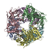 7qzfC 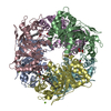 7qzgC  7qzhC 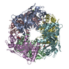 7zmjC  6yrjS S: Starting model for refinement C: citing same article ( |
|---|---|
| Similar structure data |
-Links
 PDBj
PDBj

- Assembly
| Deposited unit | 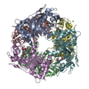
| ||||||||
|---|---|---|---|---|---|---|---|---|---|
| 1 |
| ||||||||
| Unit cell |
|
-Components
| #1: Protein | Mass: 34128.211 Da / Num. of mol.: 6 / Mutation: D152A Source method: isolated from a genetically manipulated source Source: (gene. exp.) Streptomyces lividans (bacteria) / Gene: SSPG_00656 / Production host: #2: Chemical | ChemComp-HEM /   Mass: 616.487 Da / Num. of mol.: 6 / Source method: isolated from a natural source / Formula: C34H32FeN4O4 / Feature type: SUBJECT OF INVESTIGATION Mass: 616.487 Da / Num. of mol.: 6 / Source method: isolated from a natural source / Formula: C34H32FeN4O4 / Feature type: SUBJECT OF INVESTIGATION#3: Chemical |   Mass: 24.305 Da / Num. of mol.: 3 / Source method: obtained synthetically / Formula: Mg Mass: 24.305 Da / Num. of mol.: 3 / Source method: obtained synthetically / Formula: Mg#4: Chemical | ChemComp-O /   Mass: 15.999 Da / Num. of mol.: 6 / Source method: obtained synthetically / Formula: O / Feature type: SUBJECT OF INVESTIGATION Mass: 15.999 Da / Num. of mol.: 6 / Source method: obtained synthetically / Formula: O / Feature type: SUBJECT OF INVESTIGATION#5: Water | ChemComp-HOH / |  Mass: 18.015 Da / Num. of mol.: 823 / Source method: isolated from a natural source / Formula: H2O Mass: 18.015 Da / Num. of mol.: 823 / Source method: isolated from a natural source / Formula: H2OHas ligand of interest | Y | |
|---|
-Experimental details
-Experiment
| Experiment | Method: X-RAY DIFFRACTION / Number of used crystals: 1 |
|---|
- Sample preparation
| Crystal | Density Matthews: 2.47 Å3/Da / Density % sol: 50.17 % |
|---|---|
| Crystal grow | Temperature: 293 K / Method: batch mode / pH: 7.5 Details: 6.2 mg/mL of protein in 20mM Sodium phosphate, 125mM NaCl pH 7 mixed with 125 mM MgCl2, 125 mM HEPES, 18% PEG 4000, pH 7.5 |
-Data collection
| Diffraction | Mean temperature: 301 K / Serial crystal experiment: Y |
|---|---|
| Diffraction source | Source: FREE ELECTRON LASER / Site: SACLA  / Beamline: BL2 / Wavelength: 1.127 Å / Beamline: BL2 / Wavelength: 1.127 Å |
| Detector | Type: MPCCD / Detector: CCD / Date: Jun 12, 2021 |
| Radiation | Protocol: SINGLE WAVELENGTH / Monochromatic (M) / Laue (L): M / Scattering type: x-ray |
| Radiation wavelength | Wavelength: 1.127 Å / Relative weight: 1 |
| Reflection | Resolution: 1.9→36 Å / Num. obs: 163211 / % possible obs: 100 % / Redundancy: 1510 % / CC1/2: 0.968 / R split: 0.162 / Net I/σ(I): 4.8 |
| Reflection shell | Resolution: 1.9→1.93 Å / Num. unique obs: 16133 / CC1/2: 0.282 / R split: 0.863 |
| Serial crystallography measurement | Pulse duration: 10 fsec. |
| Serial crystallography sample delivery | Description: high-viscosity cartridge-type injector / Method: injection |
| Serial crystallography sample delivery injection | Carrier solvent: hydroxyethyl cellulose / Flow rate: 0.0068 µL/min / Injector diameter: 100 µm / Power by: gas |
- Processing
| Software |
| ||||||||||||||||||||||||||||||||||||||||||||||||||||||||||||
|---|---|---|---|---|---|---|---|---|---|---|---|---|---|---|---|---|---|---|---|---|---|---|---|---|---|---|---|---|---|---|---|---|---|---|---|---|---|---|---|---|---|---|---|---|---|---|---|---|---|---|---|---|---|---|---|---|---|---|---|---|---|
| Refinement | Method to determine structure: MOLECULAR REPLACEMENT Starting model: 6YRJ Resolution: 1.9→36 Å / Cor.coef. Fo:Fc: 0.968 / Cor.coef. Fo:Fc free: 0.953 / SU B: 3.631 / SU ML: 0.1 / Cross valid method: THROUGHOUT / σ(F): 0 / ESU R: 0.128 / ESU R Free: 0.121 / Stereochemistry target values: MAXIMUM LIKELIHOOD Details: HYDROGENS HAVE BEEN ADDED IN THE RIDING POSITIONS U VALUES : REFINED INDIVIDUALLY
| ||||||||||||||||||||||||||||||||||||||||||||||||||||||||||||
| Solvent computation | Ion probe radii: 0.8 Å / Shrinkage radii: 0.8 Å / VDW probe radii: 1.2 Å / Solvent model: MASK | ||||||||||||||||||||||||||||||||||||||||||||||||||||||||||||
| Displacement parameters | Biso max: 100.24 Å2 / Biso mean: 29.268 Å2 / Biso min: 16.53 Å2
| ||||||||||||||||||||||||||||||||||||||||||||||||||||||||||||
| Refinement step | Cycle: final / Resolution: 1.9→36 Å
| ||||||||||||||||||||||||||||||||||||||||||||||||||||||||||||
| Refine LS restraints |
| ||||||||||||||||||||||||||||||||||||||||||||||||||||||||||||
| LS refinement shell | Resolution: 1.9→1.949 Å / Rfactor Rfree error: 0 / Total num. of bins used: 20
|
