 Movie
Movie Controller

[English] 日本語
 Yorodumi
- PDB-7qyh: Structure of plasmepsin II in complex with 2-aminoquinazolin-4(3H... -
+ Open data

- Basic information
| Entry | Database: PDB / ID: 7qyh | ||||||
|---|---|---|---|---|---|---|---|
| Title | Structure of plasmepsin II in complex with 2-aminoquinazolin-4(3H)-one based open-flap inhibitor | ||||||
 Components Components | Plasmepsin II | ||||||
 Keywords Keywords | HYDROLASE / Eukaryotic aspartic protease / malaria / plasmepsin | ||||||
| Function / homology |  Function and homology information Function and homology informationMHC class II antigen presentation / hemoglobin catabolic process / cytostome / plasmepsin II / vacuolar lumen / Neutrophil degranulation / food vacuole / vacuolar membrane / aspartic-type endopeptidase activity / lysosome / proteolysis Similarity search - Function | ||||||
| Biological species |  | ||||||
| Method |  X-RAY DIFFRACTION / SYNCHROTRON / MOLECULAR REPLACEMENT / Resolution: 3.33 Å X-RAY DIFFRACTION / SYNCHROTRON / MOLECULAR REPLACEMENT / Resolution: 3.33 Å | ||||||
 Authors Authors | Bobrovs, R. / Jaudzems, K. | ||||||
| Funding support | European Union, 1items
| ||||||
 Citation Citation | Journal: J.Chem.Inf.Model. / Year: 2022 Title: Exploring Aspartic Protease Inhibitor Binding to Design Selective Antimalarials. Authors: Bobrovs, R. / Basens, E.E. / Drunka, L. / Kanepe, I. / Matisone, S. / Velins, K.K. / Andrianov, V. / Leitis, G. / Zelencova-Gopejenko, D. / Rasina, D. / Jirgensons, A. / Jaudzems, K. #1: Journal: Acta Crystallogr.,Sect.D / Year: 2012 Title: Towards automated crystallographic structure refinement with phenix.refine. Authors: Afonine, P.V. / Grosse-Kunstleve, R.W. / Echols, N. / Headd, J.J. / Moriarty, N.W. / Mustyakimov, M. / Terwilliger, T.C. / Urzhumtsev, A. / Zwart, P.H. / Adams, P.D. #2: Journal: Acta Crystallogr D Struct Biol / Year: 2019 Title: Macromolecular structure determination using X-rays, neutrons and electrons: recent developments in Phenix. Authors: Dorothee Liebschner / Pavel V Afonine / Matthew L Baker / Gábor Bunkóczi / Vincent B Chen / Tristan I Croll / Bradley Hintze / Li Wei Hung / Swati Jain / Airlie J McCoy / Nigel W Moriarty ...Authors: Dorothee Liebschner / Pavel V Afonine / Matthew L Baker / Gábor Bunkóczi / Vincent B Chen / Tristan I Croll / Bradley Hintze / Li Wei Hung / Swati Jain / Airlie J McCoy / Nigel W Moriarty / Robert D Oeffner / Billy K Poon / Michael G Prisant / Randy J Read / Jane S Richardson / David C Richardson / Massimo D Sammito / Oleg V Sobolev / Duncan H Stockwell / Thomas C Terwilliger / Alexandre G Urzhumtsev / Lizbeth L Videau / Christopher J Williams / Paul D Adams /    Abstract: Diffraction (X-ray, neutron and electron) and electron cryo-microscopy are powerful methods to determine three-dimensional macromolecular structures, which are required to understand biological ...Diffraction (X-ray, neutron and electron) and electron cryo-microscopy are powerful methods to determine three-dimensional macromolecular structures, which are required to understand biological processes and to develop new therapeutics against diseases. The overall structure-solution workflow is similar for these techniques, but nuances exist because the properties of the reduced experimental data are different. Software tools for structure determination should therefore be tailored for each method. Phenix is a comprehensive software package for macromolecular structure determination that handles data from any of these techniques. Tasks performed with Phenix include data-quality assessment, map improvement, model building, the validation/rebuilding/refinement cycle and deposition. Each tool caters to the type of experimental data. The design of Phenix emphasizes the automation of procedures, where possible, to minimize repetitive and time-consuming manual tasks, while default parameters are chosen to encourage best practice. A graphical user interface provides access to many command-line features of Phenix and streamlines the transition between programs, project tracking and re-running of previous tasks. | ||||||
| History |
|
- Structure visualization
| Structure viewer | Molecule: MolmilJmol/JSmol |
|---|
- Downloads & links
-Download
| PDBx/mmCIF format | 7qyh.cif.gz | 327.7 KB | Display | PDBx/mmCIF format |
|---|---|---|---|---|
| PDB format | pdb7qyh.ent.gz | Display | PDB format | |
| PDBx/mmJSON format | 7qyh.json.gz | Tree view | PDBx/mmJSON format | |
| Others |  Other downloads Other downloads |
-Validation report
| Arichive directory | https://data.pdbj.org/pub/pdb/validation_reports/qy/7qyhftp://data.pdbj.org/pub/pdb/validation_reports/qy/7qyh | HTTPS FTP |
|---|
-Related structure data
| Related structure data | 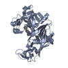 4z22S S: Starting model for refinement |
|---|---|
| Similar structure data |
-Links
 PDBj
PDBj- Assembly
| Deposited unit | 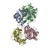
| ||||||||||||
|---|---|---|---|---|---|---|---|---|---|---|---|---|---|
| 1 | 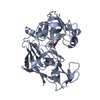
| ||||||||||||
| 2 | 
| ||||||||||||
| 3 | 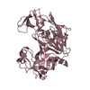
| ||||||||||||
| 4 | 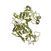
| ||||||||||||
| Unit cell |
|
-Components
| #1: Protein | Mass: 36909.617 Da / Num. of mol.: 4 Source method: isolated from a genetically manipulated source Source: (gene. exp.)  #2: Chemical | ChemComp-I0J / |   Mass: 391.506 Da / Num. of mol.: 1 / Source method: obtained synthetically / Formula: C24H29N3O2 / Feature type: SUBJECT OF INVESTIGATION Mass: 391.506 Da / Num. of mol.: 1 / Source method: obtained synthetically / Formula: C24H29N3O2 / Feature type: SUBJECT OF INVESTIGATIONHas ligand of interest | Y | Has protein modification | Y | |
|---|
-Experimental details
-Experiment
| Experiment | Method: X-RAY DIFFRACTION / Number of used crystals: 1 |
|---|
- Sample preparation
| Crystal | Density Matthews: 4.5 Å3/Da / Density % sol: 72.68 % |
|---|---|
| Crystal grow | Temperature: 298 K / Method: vapor diffusion, sitting drop / pH: 4.6 Details: 0.1M citric acid, pH 4.5, 0.3M ammonium acetate, 25% PEG 4000, protein 10 mg/mL, vapour diffusion, sitting drop, time 4-8 weeks |
-Data collection
| Diffraction | Mean temperature: 100 K / Serial crystal experiment: N |
|---|---|
| Diffraction source | Source: SYNCHROTRON / Site: SLS  / Beamline: X06DA / Wavelength: 1 Å / Beamline: X06DA / Wavelength: 1 Å |
| Detector | Type: DECTRIS PILATUS 2M-F / Detector: PIXEL / Date: Dec 9, 2021 |
| Radiation | Protocol: SINGLE WAVELENGTH / Monochromatic (M) / Laue (L): M / Scattering type: x-ray |
| Radiation wavelength | Wavelength: 1 Å / Relative weight: 1 |
| Reflection | Resolution: 3.33→137.03 Å / Num. obs: 15336 / % possible obs: 86.8 % / Redundancy: 13.1 % / Biso Wilson estimate: 68.32 Å2 / CC1/2: 0.987 / Net I/σ(I): 4.6 |
| Reflection shell | Resolution: 3.33→4.03 Å / Num. unique obs: 767 / CC1/2: 0.592 |
- Processing
| Software |
| |||||||||||||||||||||||||||||||||||||||||||||||||
|---|---|---|---|---|---|---|---|---|---|---|---|---|---|---|---|---|---|---|---|---|---|---|---|---|---|---|---|---|---|---|---|---|---|---|---|---|---|---|---|---|---|---|---|---|---|---|---|---|---|---|
| Refinement | Method to determine structure: MOLECULAR REPLACEMENT Starting model: 4Z22 Resolution: 3.33→137.03 Å / SU ML: 0.6419 / Cross valid method: FREE R-VALUE / σ(F): 1.35 / Phase error: 37.6097 Stereochemistry target values: GeoStd + Monomer Library + CDL v1.2
| |||||||||||||||||||||||||||||||||||||||||||||||||
| Solvent computation | Shrinkage radii: 0.9 Å / VDW probe radii: 1.11 Å / Solvent model: FLAT BULK SOLVENT MODEL | |||||||||||||||||||||||||||||||||||||||||||||||||
| Displacement parameters | Biso mean: 80.9 Å2 | |||||||||||||||||||||||||||||||||||||||||||||||||
| Refinement step | Cycle: LAST / Resolution: 3.33→137.03 Å
| |||||||||||||||||||||||||||||||||||||||||||||||||
| Refine LS restraints |
| |||||||||||||||||||||||||||||||||||||||||||||||||
| LS refinement shell |
|
