 Movie
Movie Controller

[English] 日本語
 Yorodumi
- PDB-7qxv: Crystal Structure of Haem-Binding Protein HemS Mutant F104A F199A... -
+ Open data

- Basic information
| Entry | Database: PDB / ID: 7qxv | ||||||
|---|---|---|---|---|---|---|---|
| Title | Crystal Structure of Haem-Binding Protein HemS Mutant F104A F199A, from Yersinia enterocolitica | ||||||
 Components Components | Hemin transport protein | ||||||
 Keywords Keywords | METAL BINDING PROTEIN / iron / haem-degrading enzyme / host-pathogen interactions | ||||||
| Function / homology | Haemin-degrading HemS/ChuX domain / Haemin-degrading HemS.ChuX domain / : / iron ion transport / DI(HYDROXYETHYL)ETHER / Hemin transport protein Function and homology information Function and homology information | ||||||
| Biological species |  Yersinia enterocolitica (bacteria) Yersinia enterocolitica (bacteria) | ||||||
| Method |  X-RAY DIFFRACTION / SYNCHROTRON / MOLECULAR REPLACEMENT / molecular replacement / Resolution: 1.67 Å X-RAY DIFFRACTION / SYNCHROTRON / MOLECULAR REPLACEMENT / molecular replacement / Resolution: 1.67 Å | ||||||
 Authors Authors | Barker, P.D. / Keith, A. / Brear, P. / Wales, D. | ||||||
| Funding support | 1items
| ||||||
 Citation Citation | Journal: To Be Published Title: Crystal Structure of Haem-Binding Protein HemS Mutant F104A F199A, from Yersinia enterocolitica Authors: Barker, P.D. / Keith, A. / Brear, P. / Wales, D. | ||||||
| History |
|
- Structure visualization
| Structure viewer | Molecule: MolmilJmol/JSmol |
|---|
- Downloads & links
-Download
| PDBx/mmCIF format | 7qxv.cif.gz | 84 KB | Display | PDBx/mmCIF format |
|---|---|---|---|---|
| PDB format | pdb7qxv.ent.gz | 61.4 KB | Display | PDB format |
| PDBx/mmJSON format | 7qxv.json.gz | Tree view | PDBx/mmJSON format | |
| Others |  Other downloads Other downloads |
-Validation report
| Summary document | 7qxv_validation.pdf.gz | 438 KB | Display | wwPDB validaton report |
|---|---|---|---|---|
| Full document | 7qxv_full_validation.pdf.gz | 440.5 KB | Display | |
| Data in XML | 7qxv_validation.xml.gz | 15 KB | Display | |
| Data in CIF | 7qxv_validation.cif.gz | 20.4 KB | Display | |
| Arichive directory | https://data.pdbj.org/pub/pdb/validation_reports/qx/7qxvftp://data.pdbj.org/pub/pdb/validation_reports/qx/7qxv | HTTPS FTP |
-Related structure data
| Related structure data | 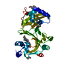 2j0pS S: Starting model for refinement |
|---|---|
| Similar structure data |
-Links
 PDBj
PDBj- Assembly
| Deposited unit | 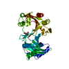
| ||||||||
|---|---|---|---|---|---|---|---|---|---|
| 1 |
| ||||||||
| Unit cell |
|
-Components
| #1: Protein | Mass: 39107.699 Da / Num. of mol.: 1 / Mutation: F104A F199A Source method: isolated from a genetically manipulated source Source: (gene. exp.) Yersinia enterocolitica (bacteria) / Gene: hemS, ERS008652_00918 / Plasmid: pET11d / Production host: |
|---|---|
| #2: Chemical | ChemComp-PEG /   Mass: 106.120 Da / Num. of mol.: 1 / Source method: obtained synthetically / Formula: C4H10O3 Mass: 106.120 Da / Num. of mol.: 1 / Source method: obtained synthetically / Formula: C4H10O3 |
| #3: Water | ChemComp-HOH /  Mass: 18.015 Da / Num. of mol.: 99 / Source method: isolated from a natural source / Formula: H2O Mass: 18.015 Da / Num. of mol.: 99 / Source method: isolated from a natural source / Formula: H2O |
| Has ligand of interest | N |
-Experimental details
-Experiment
| Experiment | Method: X-RAY DIFFRACTION / Number of used crystals: 1 |
|---|
- Sample preparation
| Crystal | Density Matthews: 2.04 Å3/Da / Density % sol: 39.59 % |
|---|---|
| Crystal grow | Temperature: 277 K / Method: vapor diffusion, hanging drop / Details: 100 mM tris-HCl 1.8 M (NH4)2SO4, 2% (w/v) PEG 400 / PH range: 8.3 |
-Data collection
| Diffraction | Mean temperature: 100 K / Serial crystal experiment: N | |||||||||||||||||||||
|---|---|---|---|---|---|---|---|---|---|---|---|---|---|---|---|---|---|---|---|---|---|---|
| Diffraction source | Source: SYNCHROTRON / Site: Diamond  / Beamline: I04 / Wavelength: 0.9793 Å / Beamline: I04 / Wavelength: 0.9793 Å | |||||||||||||||||||||
| Detector | Type: DECTRIS EIGER2 X 16M / Detector: PIXEL / Date: May 1, 2021 | |||||||||||||||||||||
| Radiation | Protocol: SINGLE WAVELENGTH / Monochromatic (M) / Laue (L): M / Scattering type: x-ray | |||||||||||||||||||||
| Radiation wavelength | Wavelength: 0.9793 Å / Relative weight: 1 | |||||||||||||||||||||
| Reflection | Resolution: 1.67→50.57 Å / Num. obs: 37692 / % possible obs: 100 % / Redundancy: 18.6 % / Rpim(I) all: 0.096 / Rrim(I) all: 0.415 / Net I/σ(I): 5.6 / Num. measured all: 701834 | |||||||||||||||||||||
| Reflection shell | Diffraction-ID: 1 / % possible all: 100
|
-Phasing
| Phasing | Method: molecular replacement |
|---|
- Processing
| Software |
| ||||||||||||||||||||||||||||||||||||||||||||||||||||||||||||
|---|---|---|---|---|---|---|---|---|---|---|---|---|---|---|---|---|---|---|---|---|---|---|---|---|---|---|---|---|---|---|---|---|---|---|---|---|---|---|---|---|---|---|---|---|---|---|---|---|---|---|---|---|---|---|---|---|---|---|---|---|---|
| Refinement | Method to determine structure: MOLECULAR REPLACEMENT Starting model: 2J0P Resolution: 1.67→50.57 Å / Cor.coef. Fo:Fc: 0.969 / Cor.coef. Fo:Fc free: 0.954 / SU B: 13.711 / SU ML: 0.322 / Cross valid method: THROUGHOUT / σ(F): 0 / ESU R: 0.152 / ESU R Free: 0.148 / Stereochemistry target values: MAXIMUM LIKELIHOOD Details: HYDROGENS HAVE BEEN ADDED IN THE RIDING POSITIONS U VALUES : REFINED INDIVIDUALLY
| ||||||||||||||||||||||||||||||||||||||||||||||||||||||||||||
| Solvent computation | Ion probe radii: 0.8 Å / Shrinkage radii: 0.8 Å / VDW probe radii: 1.2 Å / Solvent model: MASK | ||||||||||||||||||||||||||||||||||||||||||||||||||||||||||||
| Displacement parameters | Biso max: 168.68 Å2 / Biso mean: 50.554 Å2 / Biso min: 30 Å2
| ||||||||||||||||||||||||||||||||||||||||||||||||||||||||||||
| Refinement step | Cycle: final / Resolution: 1.67→50.57 Å
| ||||||||||||||||||||||||||||||||||||||||||||||||||||||||||||
| Refine LS restraints |
| ||||||||||||||||||||||||||||||||||||||||||||||||||||||||||||
| LS refinement shell | Resolution: 1.671→1.714 Å / Rfactor Rfree error: 0 / Total num. of bins used: 20
|
