 Movie
Movie Controller

[English] 日本語
 Yorodumi
- PDB-7qxc: Recognition of Staphylococcus aureus wall teichoic acid analogue ... -
+ Open data

- Basic information
| Entry | Database: PDB / ID: 7qxc | ||||||
|---|---|---|---|---|---|---|---|
| Title | Recognition of Staphylococcus aureus wall teichoic acid analogue SA533 (compound 1) by Fab4461 | ||||||
 Components Components |
| ||||||
 Keywords Keywords | CARBOHYDRATE / Antibody / Fab fragment / teichoic acid analogue | ||||||
| Function / homology | Chem-3CX / Chem-G6N / DI(HYDROXYETHYL)ETHER Function and homology information Function and homology information | ||||||
| Biological species |  Homo sapiens (human) Homo sapiens (human) | ||||||
| Method |  X-RAY DIFFRACTION / SYNCHROTRON / MOLECULAR REPLACEMENT / Resolution: 1.452 Å X-RAY DIFFRACTION / SYNCHROTRON / MOLECULAR REPLACEMENT / Resolution: 1.452 Å | ||||||
 Authors Authors | Soriano-Maldonado, P. / van Raaij, M.J. | ||||||
| Funding support |  Spain, 1items Spain, 1items
| ||||||
 Citation Citation | Journal: Acs Cent.Sci. / Year: 2022 Title: Antibody Recognition of Different Staphylococcus aureus Wall Teichoic Acid Glycoforms. Authors: Di Carluccio, C. / Soriano-Maldonado, P. / Berni, F. / de Haas, C.J.C. / Temming, A.R. / Hendriks, A. / Ali, S. / Molinaro, A. / Silipo, A. / van Sorge, N.M. / van Raaij, M.J. / Codee, J.D.C. / Marchetti, R. | ||||||
| History |
|
- Structure visualization
| Structure viewer | Molecule: MolmilJmol/JSmol |
|---|
- Downloads & links
-Download
| PDBx/mmCIF format | 7qxc.cif.gz | 116 KB | Display | PDBx/mmCIF format |
|---|---|---|---|---|
| PDB format | pdb7qxc.ent.gz | Display | PDB format | |
| PDBx/mmJSON format | 7qxc.json.gz | Tree view | PDBx/mmJSON format | |
| Others |  Other downloads Other downloads |
-Validation report
| Arichive directory | https://data.pdbj.org/pub/pdb/validation_reports/qx/7qxcftp://data.pdbj.org/pub/pdb/validation_reports/qx/7qxc | HTTPS FTP |
|---|
-Related structure data
| Related structure data | 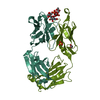 7qxdC  7qxeC 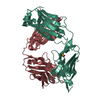 5i1dS S: Starting model for refinement C: citing same article ( |
|---|---|
| Similar structure data |
-Links
 PDBj
PDBj

- Assembly
| Deposited unit | 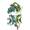
| ||||||||
|---|---|---|---|---|---|---|---|---|---|
| 1 |
| ||||||||
| Unit cell |
|
-Components
-Antibody , 2 types, 2 molecules HHHLLL
| #1: Antibody | Mass: 25147.012 Da / Num. of mol.: 1 Source method: isolated from a genetically manipulated source Source: (gene. exp.) Homo sapiens (human) / Production host:  |
|---|---|
| #2: Antibody | Mass: 24458.096 Da / Num. of mol.: 1 Source method: isolated from a genetically manipulated source Source: (gene. exp.) Homo sapiens (human) / Production host: |
-Non-polymers , 4 types, 430 molecules 


| #3: Chemical | ChemComp-3CX / ( Mass: 237.316 Da / Num. of mol.: 1 / Source method: obtained synthetically / Formula: C9H19NO4S Mass: 237.316 Da / Num. of mol.: 1 / Source method: obtained synthetically / Formula: C9H19NO4S | ||||
|---|---|---|---|---|---|
| #4: Chemical | ChemComp-PEG /  Mass: 106.120 Da / Num. of mol.: 4 / Source method: obtained synthetically / Formula: C4H10O3 Mass: 106.120 Da / Num. of mol.: 4 / Source method: obtained synthetically / Formula: C4H10O3#5: Chemical | ChemComp-G6N / [( |  Mass: 783.559 Da / Num. of mol.: 1 / Source method: obtained synthetically / Formula: C23H47NO24P2 / Feature type: SUBJECT OF INVESTIGATION Mass: 783.559 Da / Num. of mol.: 1 / Source method: obtained synthetically / Formula: C23H47NO24P2 / Feature type: SUBJECT OF INVESTIGATION#6: Water | ChemComp-HOH / | Mass: 18.015 Da / Num. of mol.: 424 / Source method: isolated from a natural source / Formula: H2O |
-Details
| Has ligand of interest | Y |
|---|---|
| Has protein modification | Y |
-Experimental details
-Experiment
| Experiment | Method: X-RAY DIFFRACTION / Number of used crystals: 1 |
|---|
- Sample preparation
| Crystal | Density Matthews: 2.8 Å3/Da / Density % sol: 56 % |
|---|---|
| Crystal grow | Temperature: 294 K / Method: vapor diffusion, sitting drop / pH: 9.5 Details: 10 mM Tris-HCl; 250 mM sodium chloride; 12.9 % w/v polyethylene glycol 20,000; 150 mM CAPSO pH 9.5; 6 % v/v polyethylene glycol 300 |
-Data collection
| Diffraction | Mean temperature: 100 K / Serial crystal experiment: N |
|---|---|
| Diffraction source | Source: SYNCHROTRON / Site: ALBA / Beamline: XALOC / Wavelength: 0.97926 Å |
| Detector | Type: DECTRIS PILATUS 6M / Detector: PIXEL / Date: Dec 1, 2020 |
| Radiation | Protocol: SINGLE WAVELENGTH / Monochromatic (M) / Laue (L): M / Scattering type: x-ray |
| Radiation wavelength | Wavelength: 0.97926 Å / Relative weight: 1 |
| Reflection | Resolution: 1.45→53.51 Å / Num. obs: 87656 / % possible obs: 96.6 % / Redundancy: 3.1 % / Biso Wilson estimate: 21.8 Å2 / CC1/2: 0.997 / Rmerge(I) obs: 0.028 / Rpim(I) all: 0.027 / Rrim(I) all: 0.039 / Net I/σ(I): 16.4 |
| Reflection shell | Resolution: 1.45→1.53 Å / Redundancy: 2.5 % / Rmerge(I) obs: 0.43 / Mean I/σ(I) obs: 1.5 / Num. unique obs: 10648 / CC1/2: 0.608 / Rpim(I) all: 0.406 / Rrim(I) all: 0.593 / % possible all: 80.6 |
- Processing
| Software |
| |||||||||||||||||||||||||||||||||||||||||||||||||||||||||||||||||||||||||||||||||||||||||||||||||||||||||||||||||||||||||||||||||||||||||||||||||||||||||||||||||||||
|---|---|---|---|---|---|---|---|---|---|---|---|---|---|---|---|---|---|---|---|---|---|---|---|---|---|---|---|---|---|---|---|---|---|---|---|---|---|---|---|---|---|---|---|---|---|---|---|---|---|---|---|---|---|---|---|---|---|---|---|---|---|---|---|---|---|---|---|---|---|---|---|---|---|---|---|---|---|---|---|---|---|---|---|---|---|---|---|---|---|---|---|---|---|---|---|---|---|---|---|---|---|---|---|---|---|---|---|---|---|---|---|---|---|---|---|---|---|---|---|---|---|---|---|---|---|---|---|---|---|---|---|---|---|---|---|---|---|---|---|---|---|---|---|---|---|---|---|---|---|---|---|---|---|---|---|---|---|---|---|---|---|---|---|---|---|---|
| Refinement | Method to determine structure: MOLECULAR REPLACEMENT Starting model: separate constant and variable domains of PDB entry 5I1D Resolution: 1.452→53.51 Å / Cor.coef. Fo:Fc: 0.972 / Cor.coef. Fo:Fc free: 0.963 / SU B: 1.231 / SU ML: 0.046 / Cross valid method: FREE R-VALUE / ESU R: 0.062 / ESU R Free: 0.064 Details: Hydrogens have been added in their riding positions
| |||||||||||||||||||||||||||||||||||||||||||||||||||||||||||||||||||||||||||||||||||||||||||||||||||||||||||||||||||||||||||||||||||||||||||||||||||||||||||||||||||||
| Solvent computation | Ion probe radii: 0.8 Å / Shrinkage radii: 0.8 Å / VDW probe radii: 1.2 Å / Solvent model: MASK BULK SOLVENT | |||||||||||||||||||||||||||||||||||||||||||||||||||||||||||||||||||||||||||||||||||||||||||||||||||||||||||||||||||||||||||||||||||||||||||||||||||||||||||||||||||||
| Displacement parameters | Biso mean: 23.016 Å2
| |||||||||||||||||||||||||||||||||||||||||||||||||||||||||||||||||||||||||||||||||||||||||||||||||||||||||||||||||||||||||||||||||||||||||||||||||||||||||||||||||||||
| Refinement step | Cycle: LAST / Resolution: 1.452→53.51 Å
| |||||||||||||||||||||||||||||||||||||||||||||||||||||||||||||||||||||||||||||||||||||||||||||||||||||||||||||||||||||||||||||||||||||||||||||||||||||||||||||||||||||
| Refine LS restraints |
| |||||||||||||||||||||||||||||||||||||||||||||||||||||||||||||||||||||||||||||||||||||||||||||||||||||||||||||||||||||||||||||||||||||||||||||||||||||||||||||||||||||
| LS refinement shell |
|
