 Movie
Movie Controller

[English] 日本語
 Yorodumi
- PDB-7qug: Crystal structure of carbon-sulfur lyase FnaPatB1 from Fusobacter... -
+ Open data

- Basic information
| Entry | Database: PDB / ID: 7qug | ||||||
|---|---|---|---|---|---|---|---|
| Title | Crystal structure of carbon-sulfur lyase FnaPatB1 from Fusobacterium nucleatum subspecies animalis in complex with allyl-cysteine | ||||||
 Components Components | carbon-sulfur lyase FnaPatB1 | ||||||
 Keywords Keywords | LYASE / carbon-sulfur lyase / PLP-dependent aminotransferase | ||||||
| Function / homology | ACETATE ION / CACODYLATE ION / allyl-cysteine Function and homology information Function and homology information | ||||||
| Biological species |  Fusobacterium nucleatum (bacteria) Fusobacterium nucleatum (bacteria) | ||||||
| Method |  X-RAY DIFFRACTION / SYNCHROTRON / MOLECULAR REPLACEMENT / Resolution: 2.6 Å X-RAY DIFFRACTION / SYNCHROTRON / MOLECULAR REPLACEMENT / Resolution: 2.6 Å | ||||||
 Authors Authors | Schwartz, M. | ||||||
| Funding support |  France, 1items France, 1items
| ||||||
 Citation Citation | Journal: J.Agric.Food Chem. / Year: 2022 Title: Metabolism of Cysteine Conjugates and Production of Flavor Sulfur Compounds by a Carbon-Sulfur Lyase from the Oral Anaerobe Fusobacterium nucleatum. Authors: Neiers, F. / Gourrat, K. / Canon, F. / Schwartz, M. | ||||||
| History |
|
- Structure visualization
| Structure viewer | Molecule: MolmilJmol/JSmol |
|---|
- Downloads & links
-Download
| PDBx/mmCIF format | 7qug.cif.gz | 204.4 KB | Display | PDBx/mmCIF format |
|---|---|---|---|---|
| PDB format | pdb7qug.ent.gz | 132.3 KB | Display | PDB format |
| PDBx/mmJSON format | 7qug.json.gz | Tree view | PDBx/mmJSON format | |
| Others |  Other downloads Other downloads |
-Validation report
| Summary document | 7qug_validation.pdf.gz | 673.1 KB | Display | wwPDB validaton report |
|---|---|---|---|---|
| Full document | 7qug_full_validation.pdf.gz | 681.4 KB | Display | |
| Data in XML | 7qug_validation.xml.gz | 27.4 KB | Display | |
| Data in CIF | 7qug_validation.cif.gz | 37.4 KB | Display | |
| Arichive directory | https://data.pdbj.org/pub/pdb/validation_reports/qu/7qugftp://data.pdbj.org/pub/pdb/validation_reports/qu/7qug | HTTPS FTP |
-Related structure data
| Related structure data |  6qp2S S: Starting model for refinement |
|---|---|
| Similar structure data |
-Links
 PDBj
PDBj


- Assembly
| Deposited unit | 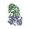
| |||||||||||||||||||||||||||||||||||||||||||||||||||||||||||||
|---|---|---|---|---|---|---|---|---|---|---|---|---|---|---|---|---|---|---|---|---|---|---|---|---|---|---|---|---|---|---|---|---|---|---|---|---|---|---|---|---|---|---|---|---|---|---|---|---|---|---|---|---|---|---|---|---|---|---|---|---|---|---|
| 1 |
| |||||||||||||||||||||||||||||||||||||||||||||||||||||||||||||
| Unit cell |
| |||||||||||||||||||||||||||||||||||||||||||||||||||||||||||||
| Noncrystallographic symmetry (NCS) | NCS domain:
NCS domain segments: Ens-ID: 1
|
-Components
| #1: Protein | Mass: 47627.426 Da / Num. of mol.: 2 Source method: isolated from a genetically manipulated source Source: (gene. exp.) Fusobacterium nucleatum (bacteria) / Production host: #2: Chemical | ChemComp-I3L / | 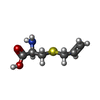  Type: L-peptide linking / Mass: 161.222 Da / Num. of mol.: 1 / Source method: obtained synthetically / Formula: C6H11NO2S / Feature type: SUBJECT OF INVESTIGATION Type: L-peptide linking / Mass: 161.222 Da / Num. of mol.: 1 / Source method: obtained synthetically / Formula: C6H11NO2S / Feature type: SUBJECT OF INVESTIGATION#3: Chemical | ChemComp-ACT / |   Mass: 59.044 Da / Num. of mol.: 1 / Source method: obtained synthetically / Formula: C2H3O2 Mass: 59.044 Da / Num. of mol.: 1 / Source method: obtained synthetically / Formula: C2H3O2#4: Chemical | ChemComp-CAC / |   Mass: 136.989 Da / Num. of mol.: 1 / Source method: obtained synthetically / Formula: C2H6AsO2 Mass: 136.989 Da / Num. of mol.: 1 / Source method: obtained synthetically / Formula: C2H6AsO2#5: Water | ChemComp-HOH / |  Mass: 18.015 Da / Num. of mol.: 10 / Source method: isolated from a natural source / Formula: H2O Mass: 18.015 Da / Num. of mol.: 10 / Source method: isolated from a natural source / Formula: H2OHas ligand of interest | Y | |
|---|
-Experimental details
-Experiment
| Experiment | Method: X-RAY DIFFRACTION / Number of used crystals: 1 |
|---|
- Sample preparation
| Crystal | Density Matthews: 3.01 Å3/Da / Density % sol: 59.16 % |
|---|---|
| Crystal grow | Temperature: 293 K / Method: vapor diffusion, sitting drop / pH: 6.5 Details: 40 % PEG 300, 0.2 M magnesium chloride in 0.1 M sodium cacodylate-HCl pH 6.5 |
-Data collection
| Diffraction | Mean temperature: 100 K / Serial crystal experiment: N |
|---|---|
| Diffraction source | Source: SYNCHROTRON / Site: ESRF / Beamline: BM07 / Wavelength: 0.979511 Å |
| Detector | Type: DECTRIS PILATUS 6M / Detector: PIXEL / Date: Oct 11, 2021 |
| Radiation | Protocol: SINGLE WAVELENGTH / Monochromatic (M) / Laue (L): M / Scattering type: x-ray |
| Radiation wavelength | Wavelength: 0.979511 Å / Relative weight: 1 |
| Reflection | Resolution: 2.6→47.43 Å / Num. obs: 36686 / % possible obs: 100 % / Redundancy: 13.1 % / Biso Wilson estimate: 59.73 Å2 / CC1/2: 1 / Rmerge(I) obs: 0.142 / Rrim(I) all: 0.148 / Net I/σ(I): 15 |
| Reflection shell | Resolution: 2.6→2.72 Å / Redundancy: 13.4 % / Rmerge(I) obs: 1.8 / Mean I/σ(I) obs: 1.6 / Num. unique obs: 4403 / CC1/2: 0.71 / % possible all: 100 |
- Processing
| Software |
| ||||||||||||||||||||||||||||||||||||||||||||||||||||||||||||||||||||||||||||||||||||||||||||||||||
|---|---|---|---|---|---|---|---|---|---|---|---|---|---|---|---|---|---|---|---|---|---|---|---|---|---|---|---|---|---|---|---|---|---|---|---|---|---|---|---|---|---|---|---|---|---|---|---|---|---|---|---|---|---|---|---|---|---|---|---|---|---|---|---|---|---|---|---|---|---|---|---|---|---|---|---|---|---|---|---|---|---|---|---|---|---|---|---|---|---|---|---|---|---|---|---|---|---|---|---|
| Refinement | Method to determine structure: MOLECULAR REPLACEMENT Starting model: 6QP2 Resolution: 2.6→19.29 Å / SU ML: 0.3497 / Cross valid method: FREE R-VALUE / σ(F): 1.34 / Phase error: 27.3827 Stereochemistry target values: GeoStd + Monomer Library + CDL v1.2
| ||||||||||||||||||||||||||||||||||||||||||||||||||||||||||||||||||||||||||||||||||||||||||||||||||
| Solvent computation | Shrinkage radii: 0.9 Å / VDW probe radii: 1.11 Å / Solvent model: FLAT BULK SOLVENT MODEL | ||||||||||||||||||||||||||||||||||||||||||||||||||||||||||||||||||||||||||||||||||||||||||||||||||
| Displacement parameters | Biso mean: 58.01 Å2 | ||||||||||||||||||||||||||||||||||||||||||||||||||||||||||||||||||||||||||||||||||||||||||||||||||
| Refinement step | Cycle: LAST / Resolution: 2.6→19.29 Å
| ||||||||||||||||||||||||||||||||||||||||||||||||||||||||||||||||||||||||||||||||||||||||||||||||||
| Refine LS restraints |
| ||||||||||||||||||||||||||||||||||||||||||||||||||||||||||||||||||||||||||||||||||||||||||||||||||
| LS refinement shell |
|
