 Movie
Movie Controller


+ Open data

- Basic information
| Entry | Database: PDB / ID: 7qta | |||||||||||||||
|---|---|---|---|---|---|---|---|---|---|---|---|---|---|---|---|---|
| Title | S1 nuclease from Aspergillus oryzae in complex with uridine | |||||||||||||||
 Components Components | Nuclease S1 | |||||||||||||||
 Keywords Keywords | HYDROLASE / Nuclease / Complex / Nucleoside / Lattice translocation defect | |||||||||||||||
| Function / homology |  Function and homology information Function and homology informationAspergillus nuclease S1 / nuclease activity / DNA catabolic process / hydrolase activity, acting on ester bonds / endonuclease activity / nucleic acid binding / metal ion binding Similarity search - Function | |||||||||||||||
| Biological species |  | |||||||||||||||
| Method |  X-RAY DIFFRACTION / SYNCHROTRON / MOLECULAR REPLACEMENT / Resolution: 1.06 Å X-RAY DIFFRACTION / SYNCHROTRON / MOLECULAR REPLACEMENT / Resolution: 1.06 Å | |||||||||||||||
 Authors Authors | Adamkova, K. / Koval, T. / Kolenko, P. / Oestergaard, L.H. / Dohnalek, J. | |||||||||||||||
| Funding support | European Union, 4items
| |||||||||||||||
 Citation Citation | Journal: Acta Crystallogr D Struct Biol / Year: 2022 Title: Atomic resolution studies of S1 nuclease complexes reveal details of RNA interaction with the enzyme despite multiple lattice-translocation defects. Authors: Adamkova, K. / Koval', T. / Ostergaard, L.H. / Duskova, J. / Maly, M. / Svecova, L. / Skalova, T. / Kolenko, P. / Dohnalek, J. | |||||||||||||||
| History |
|
- Structure visualization
| Structure viewer | Molecule: MolmilJmol/JSmol |
|---|
- Downloads & links
-Download
| PDBx/mmCIF format | 7qta.cif.gz | 254.5 KB | Display | PDBx/mmCIF format |
|---|---|---|---|---|
| PDB format | pdb7qta.ent.gz | 194.6 KB | Display | PDB format |
| PDBx/mmJSON format | 7qta.json.gz | Tree view | PDBx/mmJSON format | |
| Others |  Other downloads Other downloads |
-Validation report
| Arichive directory | https://data.pdbj.org/pub/pdb/validation_reports/qt/7qtaftp://data.pdbj.org/pub/pdb/validation_reports/qt/7qta | HTTPS FTP |
|---|
-Related structure data
| Related structure data |  7qtbC  5fb9S S: Starting model for refinement C: citing same article ( |
|---|---|
| Similar structure data |
-Links
 PDBj
PDBj

- Assembly
| Deposited unit | 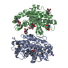
| ||||||||
|---|---|---|---|---|---|---|---|---|---|
| 1 | 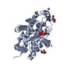
| ||||||||
| 2 | 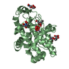
| ||||||||
| Unit cell |
|
-Components
-Protein / Sugars , 2 types, 6 molecules AB
| #1: Protein | Mass: 29083.660 Da / Num. of mol.: 2 Source method: isolated from a genetically manipulated source Source: (gene. exp.) #3: Sugar | ChemComp-NAG /  Type: D-saccharide, beta linking / Mass: 221.208 Da / Num. of mol.: 4 / Source method: obtained synthetically / Formula: C8H15NO6 Type: D-saccharide, beta linking / Mass: 221.208 Da / Num. of mol.: 4 / Source method: obtained synthetically / Formula: C8H15NO6 |
|---|
-Non-polymers , 6 types, 783 molecules 


| #2: Chemical | ChemComp-ZN /  Mass: 65.409 Da / Num. of mol.: 6 / Source method: obtained synthetically / Formula: Zn Mass: 65.409 Da / Num. of mol.: 6 / Source method: obtained synthetically / Formula: Zn#4: Chemical |  Mass: 94.971 Da / Num. of mol.: 2 / Source method: obtained synthetically / Formula: PO4 / Feature type: SUBJECT OF INVESTIGATION Mass: 94.971 Da / Num. of mol.: 2 / Source method: obtained synthetically / Formula: PO4 / Feature type: SUBJECT OF INVESTIGATION#5: Chemical |  Mass: 244.201 Da / Num. of mol.: 2 / Source method: obtained synthetically / Formula: C9H12N2O6 / Feature type: SUBJECT OF INVESTIGATION Mass: 244.201 Da / Num. of mol.: 2 / Source method: obtained synthetically / Formula: C9H12N2O6 / Feature type: SUBJECT OF INVESTIGATION#6: Chemical | ChemComp-NA / |  Mass: 22.990 Da / Num. of mol.: 1 / Source method: obtained synthetically / Formula: Na Mass: 22.990 Da / Num. of mol.: 1 / Source method: obtained synthetically / Formula: Na#7: Chemical |  Mass: 209.240 Da / Num. of mol.: 2 / Source method: obtained synthetically / Formula: C8H19NO5 / Comment: pH buffer*YM Mass: 209.240 Da / Num. of mol.: 2 / Source method: obtained synthetically / Formula: C8H19NO5 / Comment: pH buffer*YM#8: Water | ChemComp-HOH / | Mass: 18.015 Da / Num. of mol.: 770 / Source method: isolated from a natural source / Formula: H2O |
|---|
-Details
| Has ligand of interest | Y |
|---|---|
| Has protein modification | Y |
-Experimental details
-Experiment
| Experiment | Method: X-RAY DIFFRACTION / Number of used crystals: 1 |
|---|
- Sample preparation
| Crystal | Density Matthews: 1.99 Å3/Da / Density % sol: 38.16 % |
|---|---|
| Crystal grow | Temperature: 291 K / Method: vapor diffusion, sitting drop / pH: 5.5 Details: 0.2 M Sodium chloride, 0.05 M Calcium chloride, 0.1 M BIS-TRIS pH 5.5, 25% w/v Polyethylene glycol 3,350, protein concentration 10 mg/ml |
-Data collection
| Diffraction | Mean temperature: 100 K / Serial crystal experiment: N |
|---|---|
| Diffraction source | Source: SYNCHROTRON / Site: PETRA III, EMBL c/o DESY  / Beamline: P13 (MX1) / Wavelength: 1.0332 Å / Beamline: P13 (MX1) / Wavelength: 1.0332 Å |
| Detector | Type: DECTRIS PILATUS 6M / Detector: PIXEL / Date: Oct 18, 2016 |
| Radiation | Protocol: SINGLE WAVELENGTH / Monochromatic (M) / Laue (L): M / Scattering type: x-ray |
| Radiation wavelength | Wavelength: 1.0332 Å / Relative weight: 1 |
| Reflection | Resolution: 1.06→44.2 Å / Num. obs: 187610 / % possible obs: 86.02 % / Observed criterion σ(I): -3.7 / Redundancy: 3.6 % / Biso Wilson estimate: 9.8 Å2 / CC1/2: 0.997 / Rrim(I) all: 0.085 / Net I/σ(I): 8 |
| Reflection shell | Resolution: 1.06→1.088 Å / Redundancy: 3.4 % / Mean I/σ(I) obs: 1.2 / Num. unique obs: 10422 / CC1/2: 0.373 / Rrim(I) all: 1.145 / % possible all: 64.47 |
- Processing
| Software |
| ||||||||||||||||||||||||||||||||||||||||||||||||||||||||||||||||||||||||||||||||||||||||||||||||||||||||||||||||||||||||||||||||||||||||||||||||||||||
|---|---|---|---|---|---|---|---|---|---|---|---|---|---|---|---|---|---|---|---|---|---|---|---|---|---|---|---|---|---|---|---|---|---|---|---|---|---|---|---|---|---|---|---|---|---|---|---|---|---|---|---|---|---|---|---|---|---|---|---|---|---|---|---|---|---|---|---|---|---|---|---|---|---|---|---|---|---|---|---|---|---|---|---|---|---|---|---|---|---|---|---|---|---|---|---|---|---|---|---|---|---|---|---|---|---|---|---|---|---|---|---|---|---|---|---|---|---|---|---|---|---|---|---|---|---|---|---|---|---|---|---|---|---|---|---|---|---|---|---|---|---|---|---|---|---|---|---|---|---|---|---|
| Refinement | Method to determine structure: MOLECULAR REPLACEMENT Starting model: 5FB9 Resolution: 1.06→44.2 Å / Cor.coef. Fo:Fc: 0.984 / SU B: 0.684 / SU ML: 0.014 / Cross valid method: THROUGHOUT / ESU R: 0.026 Details: Hydrogens have been added in their riding positions. Structure factors were corrected for lattice translocation defect. Last refinement cycle was performed against all reflections.
| ||||||||||||||||||||||||||||||||||||||||||||||||||||||||||||||||||||||||||||||||||||||||||||||||||||||||||||||||||||||||||||||||||||||||||||||||||||||
| Solvent computation | Ion probe radii: 0.8 Å / Shrinkage radii: 0.8 Å / VDW probe radii: 1.2 Å / Solvent model: MASK BULK SOLVENT | ||||||||||||||||||||||||||||||||||||||||||||||||||||||||||||||||||||||||||||||||||||||||||||||||||||||||||||||||||||||||||||||||||||||||||||||||||||||
| Displacement parameters | Biso mean: 13.44 Å2
| ||||||||||||||||||||||||||||||||||||||||||||||||||||||||||||||||||||||||||||||||||||||||||||||||||||||||||||||||||||||||||||||||||||||||||||||||||||||
| Refinement step | Cycle: LAST / Resolution: 1.06→44.2 Å
| ||||||||||||||||||||||||||||||||||||||||||||||||||||||||||||||||||||||||||||||||||||||||||||||||||||||||||||||||||||||||||||||||||||||||||||||||||||||
| Refine LS restraints |
| ||||||||||||||||||||||||||||||||||||||||||||||||||||||||||||||||||||||||||||||||||||||||||||||||||||||||||||||||||||||||||||||||||||||||||||||||||||||
| LS refinement shell |
|
