 Movie
Movie Controller

[English] 日本語
 Yorodumi
- PDB-7qsu: Crystal structure of homing endonuclease-associated TliVMA intein... -
+ Open data

- Basic information
| Entry | Database: PDB / ID: 7qsu | |||||||||
|---|---|---|---|---|---|---|---|---|---|---|
| Title | Crystal structure of homing endonuclease-associated TliVMA intein (C1A, d333-339) | |||||||||
 Components Components | V-ATPase | |||||||||
 Keywords Keywords | HYDROLASE / intein / protein splicing / endonuclease | |||||||||
| Function / homology | Homing endonucleases / Endonuclease I-creI / Roll / Alpha Beta Function and homology information Function and homology information | |||||||||
| Biological species |   Thermococcus litoralis (archaea) Thermococcus litoralis (archaea) | |||||||||
| Method |  X-RAY DIFFRACTION / SYNCHROTRON / MOLECULAR REPLACEMENT / Resolution: 1.9 Å X-RAY DIFFRACTION / SYNCHROTRON / MOLECULAR REPLACEMENT / Resolution: 1.9 Å | |||||||||
 Authors Authors | Beyer, H.M. / Iwai, H. | |||||||||
| Funding support |  Denmark, 2items Denmark, 2items
| |||||||||
 Citation Citation | Journal: Front Mol Biosci / Year: 2022 Title: Structural Basis for the Propagation of Homing Endonuclease-Associated Inteins. Authors: Beyer, H.M. / Iwai, H. | |||||||||
| History |
|
- Structure visualization
| Structure viewer | Molecule: MolmilJmol/JSmol |
|---|
- Downloads & links
-Download
| PDBx/mmCIF format | 7qsu.cif.gz | 194.1 KB | Display | PDBx/mmCIF format |
|---|---|---|---|---|
| PDB format | pdb7qsu.ent.gz | 130.2 KB | Display | PDB format |
| PDBx/mmJSON format | 7qsu.json.gz | Tree view | PDBx/mmJSON format | |
| Others |  Other downloads Other downloads |
-Validation report
| Arichive directory | https://data.pdbj.org/pub/pdb/validation_reports/qs/7qsuftp://data.pdbj.org/pub/pdb/validation_reports/qs/7qsu | HTTPS FTP |
|---|
-Related structure data
| Related structure data |  7qssSC 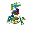 7qstC S: Starting model for refinement C: citing same article ( |
|---|---|
| Similar structure data |
-Links
 PDBj
PDBj
- Assembly
| Deposited unit | 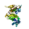
| ||||||||||||
|---|---|---|---|---|---|---|---|---|---|---|---|---|---|
| 1 |
| ||||||||||||
| Unit cell |
|
-Components
| #1: Protein | Mass: 42266.965 Da / Num. of mol.: 1 Source method: isolated from a genetically manipulated source Details: ...Details: SGKAVDGNTLVLTEEFGLVKIKELYEKLDGKGRKTVEGNEEWTELETPVTVYGYRNGRIVGIKATHIYKGISSGMIEIRTRTGRKIKVTPIHKLFTGRVTKDGLALEEVMAMHIKPGDRIAVVKKIDGGEYVKLTTSPDFRKSRKIKVPEVLDEDLAEFLGYLIADGTLKPRTVAIYNNDESLLKRANFLSTKLFGINGKIVQERTVKALLIHSKPLVDFFRKLGIPESKKARNWKVPRELLLSPPSVVKAFINAYIVCDGYYHERKGEIEITTASEEGAYGLSYLLAKLGIYATFRKKQIKGKEYYRIAISGKTNLEKLGIKRETRGYTNIDIVILFDEVVEVKYIPEPQEVYDITTETHNFVGGNMPTLLHN Source: (gene. exp.) Thermococcus litoralis (archaea) / Production host:  |
|---|---|
| #2: Chemical | ChemComp-EPE /   Mass: 238.305 Da / Num. of mol.: 1 / Source method: obtained synthetically / Formula: C8H18N2O4S / Comment: pH buffer*YM Mass: 238.305 Da / Num. of mol.: 1 / Source method: obtained synthetically / Formula: C8H18N2O4S / Comment: pH buffer*YM |
| #3: Chemical | ChemComp-MPD / (  Mass: 118.174 Da / Num. of mol.: 1 / Source method: obtained synthetically / Formula: C6H14O2 / Comment: precipitant*YM Mass: 118.174 Da / Num. of mol.: 1 / Source method: obtained synthetically / Formula: C6H14O2 / Comment: precipitant*YM |
| #4: Water | ChemComp-HOH /  Mass: 18.015 Da / Num. of mol.: 114 / Source method: isolated from a natural source / Formula: H2O Mass: 18.015 Da / Num. of mol.: 114 / Source method: isolated from a natural source / Formula: H2O |
| Has ligand of interest | N |
-Experimental details
-Experiment
| Experiment | Method: X-RAY DIFFRACTION / Number of used crystals: 1 |
|---|
- Sample preparation
| Crystal | Density Matthews: 2.52 Å3/Da / Density % sol: 51.28 % |
|---|---|
| Crystal grow | Temperature: 293 K / Method: vapor diffusion, sitting drop Details: 100 mM HEPES pH 7.5, 70% (v/v) 2-methyl-2,4-pentanediol (MPD) |
-Data collection
| Diffraction | Mean temperature: 100 K / Serial crystal experiment: N |
|---|---|
| Diffraction source | Source: SYNCHROTRON / Site: Diamond  / Beamline: I04 / Wavelength: 0.9795 Å / Beamline: I04 / Wavelength: 0.9795 Å |
| Detector | Type: DECTRIS PILATUS 6M-F / Detector: PIXEL / Date: Apr 16, 2018 |
| Radiation | Protocol: SINGLE WAVELENGTH / Monochromatic (M) / Laue (L): M / Scattering type: x-ray |
| Radiation wavelength | Wavelength: 0.9795 Å / Relative weight: 1 |
| Reflection | Resolution: 1.9→37.2 Å / Num. obs: 33640 / % possible obs: 99.8 % / Redundancy: 6.6 % / Biso Wilson estimate: 38.75 Å2 / CC1/2: 1 / Rmerge(I) obs: 0.858 / Net I/σ(I): 19.3 |
| Reflection shell | Resolution: 1.9→2.01 Å / Num. unique obs: 5361 / CC1/2: 0.793 |
- Processing
| Software |
| ||||||||||||||||||||||||||||||||||||||||||||||||||||||||||||||||||||||||||||||||||||||||||||||||||||||||||||||||||||||||||||||||||||||||||||||||||||||
|---|---|---|---|---|---|---|---|---|---|---|---|---|---|---|---|---|---|---|---|---|---|---|---|---|---|---|---|---|---|---|---|---|---|---|---|---|---|---|---|---|---|---|---|---|---|---|---|---|---|---|---|---|---|---|---|---|---|---|---|---|---|---|---|---|---|---|---|---|---|---|---|---|---|---|---|---|---|---|---|---|---|---|---|---|---|---|---|---|---|---|---|---|---|---|---|---|---|---|---|---|---|---|---|---|---|---|---|---|---|---|---|---|---|---|---|---|---|---|---|---|---|---|---|---|---|---|---|---|---|---|---|---|---|---|---|---|---|---|---|---|---|---|---|---|---|---|---|---|---|---|---|
| Refinement | Method to determine structure: MOLECULAR REPLACEMENT Starting model: 7QSS Resolution: 1.9→37.2 Å / SU ML: 0.2023 / Cross valid method: FREE R-VALUE / σ(F): 1.35 / Phase error: 22.1656 Stereochemistry target values: GeoStd + Monomer Library + CDL v1.2
| ||||||||||||||||||||||||||||||||||||||||||||||||||||||||||||||||||||||||||||||||||||||||||||||||||||||||||||||||||||||||||||||||||||||||||||||||||||||
| Solvent computation | Shrinkage radii: 0.9 Å / VDW probe radii: 1.11 Å / Solvent model: FLAT BULK SOLVENT MODEL | ||||||||||||||||||||||||||||||||||||||||||||||||||||||||||||||||||||||||||||||||||||||||||||||||||||||||||||||||||||||||||||||||||||||||||||||||||||||
| Displacement parameters | Biso mean: 50.34 Å2 | ||||||||||||||||||||||||||||||||||||||||||||||||||||||||||||||||||||||||||||||||||||||||||||||||||||||||||||||||||||||||||||||||||||||||||||||||||||||
| Refinement step | Cycle: LAST / Resolution: 1.9→37.2 Å
| ||||||||||||||||||||||||||||||||||||||||||||||||||||||||||||||||||||||||||||||||||||||||||||||||||||||||||||||||||||||||||||||||||||||||||||||||||||||
| Refine LS restraints |
| ||||||||||||||||||||||||||||||||||||||||||||||||||||||||||||||||||||||||||||||||||||||||||||||||||||||||||||||||||||||||||||||||||||||||||||||||||||||
| LS refinement shell |
| ||||||||||||||||||||||||||||||||||||||||||||||||||||||||||||||||||||||||||||||||||||||||||||||||||||||||||||||||||||||||||||||||||||||||||||||||||||||
| Refinement TLS params. | Method: refined / Refine-ID: X-RAY DIFFRACTION
| ||||||||||||||||||||||||||||||||||||||||||||||||||||||||||||||||||||||||||||||||||||||||||||||||||||||||||||||||||||||||||||||||||||||||||||||||||||||
| Refinement TLS group |
|
