 Movie
Movie Controller

[English] 日本語
 Yorodumi
- PDB-7qsp: Permutated C-terminal lobe of the ribose binding protein from The... -
+ Open data

- Basic information
| Entry | Database: PDB / ID: 7qsp | ||||||
|---|---|---|---|---|---|---|---|
| Title | Permutated C-terminal lobe of the ribose binding protein from Thermotoga maritima | ||||||
 Components Components | Ribose ABC transporter, periplasmic ribose-binding protein | ||||||
 Keywords Keywords | SUGAR BINDING PROTEIN / Solute binding protein / circular permutation / Periplasmic binding proteins / domain swapping | ||||||
| Function / homology |  Function and homology information Function and homology informationmonosaccharide binding / transmembrane transport / outer membrane-bounded periplasmic space Similarity search - Function | ||||||
| Biological species |   Thermotoga maritima (bacteria) Thermotoga maritima (bacteria) | ||||||
| Method |  X-RAY DIFFRACTION / SYNCHROTRON / MOLECULAR REPLACEMENT / Resolution: 1.36 Å X-RAY DIFFRACTION / SYNCHROTRON / MOLECULAR REPLACEMENT / Resolution: 1.36 Å | ||||||
 Authors Authors | Shanmugaratnam, S. / Michel, F. / Hocker, B. | ||||||
| Funding support | European Union, 1items
| ||||||
 Citation Citation | Journal: Acta Crystallogr D Struct Biol / Year: 2023 Title: Structures of permuted halves of a modern ribose-binding protein. Authors: Michel, F. / Shanmugaratnam, S. / Romero-Romero, S. / Hocker, B. | ||||||
| History |
|
- Structure visualization
| Structure viewer | Molecule: MolmilJmol/JSmol |
|---|
- Downloads & links
-Download
| PDBx/mmCIF format | 7qsp.cif.gz | 196.7 KB | Display | PDBx/mmCIF format |
|---|---|---|---|---|
| PDB format | pdb7qsp.ent.gz | 132.1 KB | Display | PDB format |
| PDBx/mmJSON format | 7qsp.json.gz | Tree view | PDBx/mmJSON format | |
| Others |  Other downloads Other downloads |
-Validation report
| Arichive directory | https://data.pdbj.org/pub/pdb/validation_reports/qs/7qspftp://data.pdbj.org/pub/pdb/validation_reports/qs/7qsp | HTTPS FTP |
|---|
-Related structure data
| Related structure data | 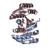 7qsqC  2fn9S S: Starting model for refinement C: citing same article ( |
|---|---|
| Similar structure data |
-Links
 PDBj
PDBj
- Assembly
| Deposited unit | 
| ||||||||||||
|---|---|---|---|---|---|---|---|---|---|---|---|---|---|
| 1 |
| ||||||||||||
| Unit cell |
|
-Components
| #1: Protein | Mass: 16932.090 Da / Num. of mol.: 2 Source method: isolated from a genetically manipulated source Source: (gene. exp.) Thermotoga maritima (bacteria) / Gene: TM_0958 / Production host: #2: Chemical | ChemComp-EDO /   Mass: 62.068 Da / Num. of mol.: 9 / Source method: obtained synthetically / Formula: C2H6O2 Mass: 62.068 Da / Num. of mol.: 9 / Source method: obtained synthetically / Formula: C2H6O2#3: Water | ChemComp-HOH / |  Mass: 18.015 Da / Num. of mol.: 196 / Source method: isolated from a natural source / Formula: H2O Mass: 18.015 Da / Num. of mol.: 196 / Source method: isolated from a natural source / Formula: H2OHas ligand of interest | N | |
|---|
-Experimental details
-Experiment
| Experiment | Method: X-RAY DIFFRACTION / Number of used crystals: 1 |
|---|
- Sample preparation
| Crystal | Density % sol: 27.97 % |
|---|---|
| Crystal grow | Temperature: 293 K / Method: vapor diffusion, sitting drop Details: 0.2 M Magnesium acetate, 20% (w/v) PEG3350, cryogenic additives: 20% PEG3000, 20% Ethylene-glycol, 0.2M KNO3 |
-Data collection
| Diffraction | Mean temperature: 100 K / Serial crystal experiment: N |
|---|---|
| Diffraction source | Source: SYNCHROTRON / Site: BESSY  / Beamline: 14.1 / Wavelength: 0.9184 Å / Beamline: 14.1 / Wavelength: 0.9184 Å |
| Detector | Type: DECTRIS PILATUS 6M / Detector: PIXEL / Date: Apr 25, 2019 |
| Radiation | Monochromator: DCM Si(111) / Protocol: SINGLE WAVELENGTH / Monochromatic (M) / Laue (L): M / Scattering type: x-ray |
| Radiation wavelength | Wavelength: 0.9184 Å / Relative weight: 1 |
| Reflection | Resolution: 1.36→39.76 Å / Num. obs: 50882 / % possible obs: 99 % / Redundancy: 10.5 % / Biso Wilson estimate: 18.76 Å2 / CC1/2: 0.999 / CC star: 1 / Rmerge(I) obs: 0.081 / Rpim(I) all: 0.026 / Rrim(I) all: 0.085 / Net I/σ(I): 13.93 |
| Reflection shell | Resolution: 1.36→1.4 Å / Redundancy: 9.9 % / Rmerge(I) obs: 1.907 / Mean I/σ(I) obs: 1 / Num. unique obs: 4875 / CC1/2: 0.322 / CC star: 0.698 / Rpim(I) all: 0.616 / Rrim(I) all: 2.008 / % possible all: 96.7 |
- Processing
| Software |
| ||||||||||||||||||||||||||||||||||||||||||||||||||||||||||||||||||||||||||||||||||||||||||||||||||||||||||||||||
|---|---|---|---|---|---|---|---|---|---|---|---|---|---|---|---|---|---|---|---|---|---|---|---|---|---|---|---|---|---|---|---|---|---|---|---|---|---|---|---|---|---|---|---|---|---|---|---|---|---|---|---|---|---|---|---|---|---|---|---|---|---|---|---|---|---|---|---|---|---|---|---|---|---|---|---|---|---|---|---|---|---|---|---|---|---|---|---|---|---|---|---|---|---|---|---|---|---|---|---|---|---|---|---|---|---|---|---|---|---|---|---|---|---|
| Refinement | Method to determine structure: MOLECULAR REPLACEMENT Starting model: 2FN9 Resolution: 1.36→39.76 Å / SU ML: 0.1974 / Cross valid method: FREE R-VALUE / Phase error: 19.3755 Stereochemistry target values: GeoStd + Monomer Library + CDL v1.2
| ||||||||||||||||||||||||||||||||||||||||||||||||||||||||||||||||||||||||||||||||||||||||||||||||||||||||||||||||
| Solvent computation | Shrinkage radii: 0.9 Å / VDW probe radii: 1.11 Å / Solvent model: FLAT BULK SOLVENT MODEL | ||||||||||||||||||||||||||||||||||||||||||||||||||||||||||||||||||||||||||||||||||||||||||||||||||||||||||||||||
| Displacement parameters | Biso mean: 25.58 Å2 | ||||||||||||||||||||||||||||||||||||||||||||||||||||||||||||||||||||||||||||||||||||||||||||||||||||||||||||||||
| Refinement step | Cycle: LAST / Resolution: 1.36→39.76 Å
| ||||||||||||||||||||||||||||||||||||||||||||||||||||||||||||||||||||||||||||||||||||||||||||||||||||||||||||||||
| Refine LS restraints |
| ||||||||||||||||||||||||||||||||||||||||||||||||||||||||||||||||||||||||||||||||||||||||||||||||||||||||||||||||
| LS refinement shell |
| ||||||||||||||||||||||||||||||||||||||||||||||||||||||||||||||||||||||||||||||||||||||||||||||||||||||||||||||||
| Refinement TLS params. | Method: refined / Refine-ID: X-RAY DIFFRACTION
| ||||||||||||||||||||||||||||||||||||||||||||||||||||||||||||||||||||||||||||||||||||||||||||||||||||||||||||||||
| Refinement TLS group | Refine-ID: X-RAY DIFFRACTION / Auth seq-ID: 1 - 142
|
