 Movie
Movie Controller


+ Open data

- Basic information
| Entry | Database: PDB / ID: 7qrj | ||||||
|---|---|---|---|---|---|---|---|
| Title | Crystal structure of Zamilon vitis protein Zav_19 | ||||||
 Components Components | Zav_19 protein | ||||||
 Keywords Keywords | VIRAL PROTEIN / Receptor Binding Protein Fiber head | ||||||
| Function / homology | Minor virion protein Function and homology information Function and homology information | ||||||
| Biological species |  Zamilon virus Zamilon virus | ||||||
| Method |  X-RAY DIFFRACTION / SYNCHROTRON / SAD / Resolution: 1.38 Å X-RAY DIFFRACTION / SYNCHROTRON / SAD / Resolution: 1.38 Å | ||||||
 Authors Authors | Jeudy, S. / Abergel, C. | ||||||
| Funding support |  France, 1items France, 1items
| ||||||
 Citation Citation | Journal: To Be Published Title: The fibre head structure used by unrelated families of viruses is unexpectedly a major component of the Marseilleviridae and Zamilon virophages capsids Authors: Jeudy, S. / Abergel, C. | ||||||
| History |
|
- Structure visualization
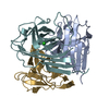
| Structure viewer | Molecule: MolmilJmol/JSmol |
|---|
- Downloads & links
-Download
| PDBx/mmCIF format | 7qrj.cif.gz | 458.6 KB | Display | PDBx/mmCIF format |
|---|---|---|---|---|
| PDB format | pdb7qrj.ent.gz | 378.8 KB | Display | PDB format |
| PDBx/mmJSON format | 7qrj.json.gz | Tree view | PDBx/mmJSON format | |
| Others |  Other downloads Other downloads |
-Validation report
| Arichive directory | https://data.pdbj.org/pub/pdb/validation_reports/qr/7qrjftp://data.pdbj.org/pub/pdb/validation_reports/qr/7qrj | HTTPS FTP |
|---|
-Related structure data
-Links
 PDBj
PDBj- Assembly
| Deposited unit | 
| ||||||||
|---|---|---|---|---|---|---|---|---|---|
| 1 | 
| ||||||||
| 2 | 
| ||||||||
| Unit cell |
|
-Components
| #1: Protein | Mass: 19601.871 Da / Num. of mol.: 6 Source method: isolated from a genetically manipulated source Source: (gene. exp.) Zamilon virus / Gene: za3_19 / Plasmid: pET-Duet / Production host:  #2: Water | ChemComp-HOH / |  Mass: 18.015 Da / Num. of mol.: 1438 / Source method: isolated from a natural source / Formula: H2O Mass: 18.015 Da / Num. of mol.: 1438 / Source method: isolated from a natural source / Formula: H2O |
|---|
-Experimental details
-Experiment
| Experiment | Method: X-RAY DIFFRACTION / Number of used crystals: 1 |
|---|
- Sample preparation
| Crystal | Density Matthews: 2.6 Å3/Da / Density % sol: 52.63 % |
|---|---|
| Crystal grow | Temperature: 293 K / Method: vapor diffusion, hanging drop / pH: 4 / Details: PEG 3350, ammonium citrate |
-Data collection
| Diffraction | Mean temperature: 100 K / Serial crystal experiment: N | ||||||||||||||||||||||||||||||
|---|---|---|---|---|---|---|---|---|---|---|---|---|---|---|---|---|---|---|---|---|---|---|---|---|---|---|---|---|---|---|---|
| Diffraction source | Source: SYNCHROTRON / Site: SOLEIL / Beamline: PROXIMA 1 / Wavelength: 0.9793 Å | ||||||||||||||||||||||||||||||
| Detector | Type: DECTRIS PILATUS 6M / Detector: PIXEL / Date: Jan 30, 2018 | ||||||||||||||||||||||||||||||
| Radiation | Protocol: SINGLE WAVELENGTH / Monochromatic (M) / Laue (L): M / Scattering type: x-ray | ||||||||||||||||||||||||||||||
| Radiation wavelength | Wavelength: 0.9793 Å / Relative weight: 1 | ||||||||||||||||||||||||||||||
| Reflection | Resolution: 1.38→45.25 Å / Num. obs: 245617 / % possible obs: 99.9 % / Redundancy: 4.8 % / CC1/2: 0.994 / Rmerge(I) obs: 0.057 / Rpim(I) all: 0.029 / Rrim(I) all: 0.064 / Net I/σ(I): 13.7 / Num. measured all: 1177549 / Scaling rejects: 40662 | ||||||||||||||||||||||||||||||
| Reflection shell | Diffraction-ID: 1
|
-Phasing
| Phasing | Method: SAD |
|---|
- Processing
| Software |
| ||||||||||||||||||||
|---|---|---|---|---|---|---|---|---|---|---|---|---|---|---|---|---|---|---|---|---|---|
| Refinement | Method to determine structure: SAD / Resolution: 1.38→45.25 Å / Cross valid method: THROUGHOUT
| ||||||||||||||||||||
| Displacement parameters | Biso max: 56.08 Å2 / Biso mean: 16.019 Å2 / Biso min: 5.97 Å2 | ||||||||||||||||||||
| Refinement step | Cycle: LAST / Resolution: 1.38→45.25 Å
| ||||||||||||||||||||
| LS refinement shell | Resolution: 1.38→1.4 Å
|
