 Movie
Movie Controller


+ Open data

- Basic information
| Entry | Database: PDB / ID: 7qqi | ||||||
|---|---|---|---|---|---|---|---|
| Title | Sucrose phosphorylase from Faecalibaculum rodentium | ||||||
 Components Components | Aamy domain-containing protein | ||||||
 Keywords Keywords | TRANSFERASE / sucrose phosphorylase | ||||||
| Function / homology |  Function and homology information Function and homology information1,4-alpha-oligoglucan phosphorylase activity / carbohydrate metabolic process Similarity search - Function | ||||||
| Biological species |  Faecalibaculum rodentium (bacteria) Faecalibaculum rodentium (bacteria) | ||||||
| Method |  X-RAY DIFFRACTION / SYNCHROTRON / MOLECULAR REPLACEMENT / Resolution: 1.36 Å X-RAY DIFFRACTION / SYNCHROTRON / MOLECULAR REPLACEMENT / Resolution: 1.36 Å | ||||||
 Authors Authors | Ubiparip, Z. / Capra, N. / Rozeboom, H.J. / Desmet, T. / Thunnissen, A.M.W.H. | ||||||
| Funding support | European Union, 1items
| ||||||
 Citation Citation | Journal: To Be Published Title: Sucrose phosphorylase from Faecalibaculum rodentium Authors: Ubiparip, Z. / Capra, N. / Rozeboom, H.J. / Thunnissen, A.M.W.H. / Desmet, T. | ||||||
| History |
|
- Structure visualization
| Structure viewer | Molecule: MolmilJmol/JSmol |
|---|
- Downloads & links
-Download
| PDBx/mmCIF format | 7qqi.cif.gz | 365 KB | Display | PDBx/mmCIF format |
|---|---|---|---|---|
| PDB format | pdb7qqi.ent.gz | 287.6 KB | Display | PDB format |
| PDBx/mmJSON format | 7qqi.json.gz | Tree view | PDBx/mmJSON format | |
| Others |  Other downloads Other downloads |
-Validation report
| Arichive directory | https://data.pdbj.org/pub/pdb/validation_reports/qq/7qqiftp://data.pdbj.org/pub/pdb/validation_reports/qq/7qqi | HTTPS FTP |
|---|
-Related structure data
| Related structure data |  6s9vS S: Starting model for refinement |
|---|---|
| Similar structure data |
-Links
 PDBj
PDBj

- Assembly
| Deposited unit | 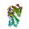
| ||||||||
|---|---|---|---|---|---|---|---|---|---|
| 1 |
| ||||||||
| Unit cell |
|
-Components
| #1: Protein | Mass: 56898.738 Da / Num. of mol.: 1 Source method: isolated from a genetically manipulated source Source: (gene. exp.) Faecalibaculum rodentium (bacteria) / Gene: AALO17_25870 / Production host: |
|---|---|
| #2: Chemical | ChemComp-TRS /   Mass: 122.143 Da / Num. of mol.: 1 / Source method: obtained synthetically / Formula: C4H12NO3 / Comment: pH buffer*YM Mass: 122.143 Da / Num. of mol.: 1 / Source method: obtained synthetically / Formula: C4H12NO3 / Comment: pH buffer*YM |
| #3: Water | ChemComp-HOH /  Mass: 18.015 Da / Num. of mol.: 448 / Source method: isolated from a natural source / Formula: H2O Mass: 18.015 Da / Num. of mol.: 448 / Source method: isolated from a natural source / Formula: H2O |
| Has ligand of interest | N |
-Experimental details
-Experiment
| Experiment | Method: X-RAY DIFFRACTION / Number of used crystals: 1 |
|---|
- Sample preparation
| Crystal | Density Matthews: 1.95 Å3/Da / Density % sol: 37 % |
|---|---|
| Crystal grow | Temperature: 291 K / Method: vapor diffusion, sitting drop / pH: 7 Details: Protein was concentrated to 30 mg/ml in 25 mM Tris, pH 7.8, 100 mM NaCl. Drops were prepared by mixing protein and reservoir solution at a 1:1 volume ratio. Composition reservoir solution: 1 ...Details: Protein was concentrated to 30 mg/ml in 25 mM Tris, pH 7.8, 100 mM NaCl. Drops were prepared by mixing protein and reservoir solution at a 1:1 volume ratio. Composition reservoir solution: 1 M Na citrate, 0.1 M Tris, pH 7.0, 0.2 M NaCl |
-Data collection
| Diffraction | Mean temperature: 100 K / Serial crystal experiment: N | |||||||||||||||||||||
|---|---|---|---|---|---|---|---|---|---|---|---|---|---|---|---|---|---|---|---|---|---|---|
| Diffraction source | Source: SYNCHROTRON / Site: ESRF  / Beamline: MASSIF-1 / Wavelength: 0.965459 Å / Beamline: MASSIF-1 / Wavelength: 0.965459 Å | |||||||||||||||||||||
| Detector | Type: DECTRIS PILATUS3 2M / Detector: PIXEL / Date: Nov 25, 2020 | |||||||||||||||||||||
| Radiation | Protocol: SINGLE WAVELENGTH / Monochromatic (M) / Laue (L): M / Scattering type: x-ray | |||||||||||||||||||||
| Radiation wavelength | Wavelength: 0.965459 Å / Relative weight: 1 | |||||||||||||||||||||
| Reflection | Resolution: 1.36→46 Å / Num. obs: 95834 / % possible obs: 99.7 % / Redundancy: 5 % / CC1/2: 0.999 / Rmerge(I) obs: 0.076 / Rpim(I) all: 0.056 / Rrim(I) all: 0.095 / Net I/σ(I): 10.7 | |||||||||||||||||||||
| Reflection shell | Diffraction-ID: 1
|
- Processing
| Software |
| ||||||||||||||||||||||||||||||||||||||||||||||||||||||||||||||||||||||||||||||||||||||||||||||||||||||||||||||||||||||||||||||||||||||||||||||||||||||||||||||||
|---|---|---|---|---|---|---|---|---|---|---|---|---|---|---|---|---|---|---|---|---|---|---|---|---|---|---|---|---|---|---|---|---|---|---|---|---|---|---|---|---|---|---|---|---|---|---|---|---|---|---|---|---|---|---|---|---|---|---|---|---|---|---|---|---|---|---|---|---|---|---|---|---|---|---|---|---|---|---|---|---|---|---|---|---|---|---|---|---|---|---|---|---|---|---|---|---|---|---|---|---|---|---|---|---|---|---|---|---|---|---|---|---|---|---|---|---|---|---|---|---|---|---|---|---|---|---|---|---|---|---|---|---|---|---|---|---|---|---|---|---|---|---|---|---|---|---|---|---|---|---|---|---|---|---|---|---|---|---|---|---|---|
| Refinement | Method to determine structure: MOLECULAR REPLACEMENT Starting model: 6S9V Resolution: 1.36→46 Å / Cor.coef. Fo:Fc: 0.978 / Cor.coef. Fo:Fc free: 0.969 / SU B: 3.355 / SU ML: 0.056 / Cross valid method: FREE R-VALUE / ESU R: 0.061 / ESU R Free: 0.057 Details: Hydrogens have been added in their riding positions
| ||||||||||||||||||||||||||||||||||||||||||||||||||||||||||||||||||||||||||||||||||||||||||||||||||||||||||||||||||||||||||||||||||||||||||||||||||||||||||||||||
| Solvent computation | Ion probe radii: 0.8 Å / Shrinkage radii: 0.8 Å / VDW probe radii: 1.2 Å / Solvent model: MASK BULK SOLVENT | ||||||||||||||||||||||||||||||||||||||||||||||||||||||||||||||||||||||||||||||||||||||||||||||||||||||||||||||||||||||||||||||||||||||||||||||||||||||||||||||||
| Displacement parameters | Biso mean: 16.554 Å2
| ||||||||||||||||||||||||||||||||||||||||||||||||||||||||||||||||||||||||||||||||||||||||||||||||||||||||||||||||||||||||||||||||||||||||||||||||||||||||||||||||
| Refinement step | Cycle: LAST / Resolution: 1.36→46 Å
| ||||||||||||||||||||||||||||||||||||||||||||||||||||||||||||||||||||||||||||||||||||||||||||||||||||||||||||||||||||||||||||||||||||||||||||||||||||||||||||||||
| Refine LS restraints |
| ||||||||||||||||||||||||||||||||||||||||||||||||||||||||||||||||||||||||||||||||||||||||||||||||||||||||||||||||||||||||||||||||||||||||||||||||||||||||||||||||
| LS refinement shell |
|
