 Movie
Movie Controller

[English] 日本語
 Yorodumi
- PDB-7qq0: X-ray structure of the adduct obtained upon reaction of [cis-Rh2(... -
+ Open data

- Basic information
| Entry | Database: PDB / ID: 7qq0 | ||||||
|---|---|---|---|---|---|---|---|
| Title | X-ray structure of the adduct obtained upon reaction of [cis-Rh2(OCOCH3)2(OCOCF3)2] with RNase A (2) | ||||||
 Components Components | Ribonuclease pancreatic | ||||||
 Keywords Keywords | HYDROLASE / Protein metalation / dirhodium tetraacetate derivative / dimetallic paddlewheel complexes | ||||||
| Function / homology |  Function and homology information Function and homology informationpancreatic ribonuclease / ribonuclease A activity / RNA nuclease activity / nucleic acid binding / defense response to Gram-positive bacterium / hydrolase activity / extracellular region Similarity search - Function | ||||||
| Biological species |  | ||||||
| Method |  X-RAY DIFFRACTION / SYNCHROTRON / MOLECULAR REPLACEMENT / Resolution: 1.32 Å X-RAY DIFFRACTION / SYNCHROTRON / MOLECULAR REPLACEMENT / Resolution: 1.32 Å | ||||||
 Authors Authors | Loreto, D. / Merlino, A. | ||||||
| Funding support |  Italy, 1items Italy, 1items
| ||||||
 Citation Citation | Journal: Dalton Trans / Year: 2022 Title: Reactivity of a fluorine-containing dirhodium tetracarboxylate compound with proteins. Authors: Loreto, D. / Esposito, A. / Demitri, N. / Guaragna, A. / Merlino, A. | ||||||
| History |
|
- Structure visualization
| Structure viewer | Molecule: MolmilJmol/JSmol |
|---|
- Downloads & links
-Download
| PDBx/mmCIF format | 7qq0.cif.gz | 78.1 KB | Display | PDBx/mmCIF format |
|---|---|---|---|---|
| PDB format | pdb7qq0.ent.gz | Display | PDB format | |
| PDBx/mmJSON format | 7qq0.json.gz | Tree view | PDBx/mmJSON format | |
| Others |  Other downloads Other downloads |
-Validation report
| Arichive directory | https://data.pdbj.org/pub/pdb/validation_reports/qq/7qq0ftp://data.pdbj.org/pub/pdb/validation_reports/qq/7qq0 | HTTPS FTP |
|---|
-Related structure data
| Related structure data |  7qpwC  7qpyC  7qpzC 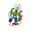 7qq1C  1jvtS S: Starting model for refinement C: citing same article ( |
|---|---|
| Similar structure data |
-Links
 PDBj
PDBj

- Assembly
| Deposited unit | 
| ||||||||
|---|---|---|---|---|---|---|---|---|---|
| 1 | 
| ||||||||
| 2 | 
| ||||||||
| Unit cell |
| ||||||||
| Components on special symmetry positions |
|
-Components
| #1: Protein | Mass: 13708.326 Da / Num. of mol.: 2 / Source method: isolated from a natural source / Source: (natural) #2: Chemical |   Mass: 367.907 Da / Num. of mol.: 2 / Source method: obtained synthetically / Formula: C2H10O8Rh2 / Feature type: SUBJECT OF INVESTIGATION Mass: 367.907 Da / Num. of mol.: 2 / Source method: obtained synthetically / Formula: C2H10O8Rh2 / Feature type: SUBJECT OF INVESTIGATION#3: Chemical |   Mass: 102.906 Da / Num. of mol.: 2 / Source method: obtained synthetically / Formula: Rh Mass: 102.906 Da / Num. of mol.: 2 / Source method: obtained synthetically / Formula: Rh#4: Water | ChemComp-HOH / |  Mass: 18.015 Da / Num. of mol.: 387 / Source method: isolated from a natural source / Formula: H2O Mass: 18.015 Da / Num. of mol.: 387 / Source method: isolated from a natural source / Formula: H2OHas ligand of interest | Y | Has protein modification | Y | |
|---|
-Experimental details
-Experiment
| Experiment | Method: X-RAY DIFFRACTION / Number of used crystals: 1 |
|---|
- Sample preparation
| Crystal |
| |||||||||
|---|---|---|---|---|---|---|---|---|---|---|
| Crystal grow | Temperature: 298 K / Method: vapor diffusion, hanging drop / pH: 5.1 / Details: 22% PEG 4K 10 mM sodium citrate buffer pH 5.1 |
-Data collection
| Diffraction | Mean temperature: 100 K / Serial crystal experiment: N |
|---|---|
| Diffraction source | Source: SYNCHROTRON / Site: ELETTRA / Beamline: 11.2C / Wavelength: 1 Å |
| Detector | Type: DECTRIS PILATUS 6M / Detector: PIXEL / Date: May 22, 2021 |
| Radiation | Protocol: SINGLE WAVELENGTH / Monochromatic (M) / Laue (L): M / Scattering type: x-ray |
| Radiation wavelength | Wavelength: 1 Å / Relative weight: 1 |
| Reflection | Resolution: 1.32→41.143 Å / Num. obs: 54908 / % possible obs: 99.5 % / Redundancy: 5.5 % / CC1/2: 0.996 / Rmerge(I) obs: 0.138 / Rpim(I) all: 0.1 / Net I/σ(I): 10.8 |
| Reflection shell | Resolution: 1.32→1.35 Å / Redundancy: 5.8 % / Rmerge(I) obs: 0.862 / Mean I/σ(I) obs: 2.2 / Num. unique obs: 2752 / CC1/2: 0.704 / Rpim(I) all: 0.57 / % possible all: 100 |
- Processing
| Software |
| |||||||||||||||||||||||||||||||||||||||||||||||||||||||||||||||||||||||||||||||||||||||||||||||||||||||||||||||||||||||||||||||||||||||||||||||||||||||||||
|---|---|---|---|---|---|---|---|---|---|---|---|---|---|---|---|---|---|---|---|---|---|---|---|---|---|---|---|---|---|---|---|---|---|---|---|---|---|---|---|---|---|---|---|---|---|---|---|---|---|---|---|---|---|---|---|---|---|---|---|---|---|---|---|---|---|---|---|---|---|---|---|---|---|---|---|---|---|---|---|---|---|---|---|---|---|---|---|---|---|---|---|---|---|---|---|---|---|---|---|---|---|---|---|---|---|---|---|---|---|---|---|---|---|---|---|---|---|---|---|---|---|---|---|---|---|---|---|---|---|---|---|---|---|---|---|---|---|---|---|---|---|---|---|---|---|---|---|---|---|---|---|---|---|---|---|---|
| Refinement | Method to determine structure: MOLECULAR REPLACEMENT Starting model: 1JVT Resolution: 1.32→41.14 Å / Cor.coef. Fo:Fc: 0.938 / Cor.coef. Fo:Fc free: 0.89 / SU B: 1.088 / SU ML: 0.047 / Cross valid method: FREE R-VALUE / ESU R: 0.067 / ESU R Free: 0.07 Details: Hydrogens have been added in their riding positions
| |||||||||||||||||||||||||||||||||||||||||||||||||||||||||||||||||||||||||||||||||||||||||||||||||||||||||||||||||||||||||||||||||||||||||||||||||||||||||||
| Solvent computation | Ion probe radii: 0.8 Å / Shrinkage radii: 0.8 Å / VDW probe radii: 1.2 Å / Solvent model: MASK BULK SOLVENT | |||||||||||||||||||||||||||||||||||||||||||||||||||||||||||||||||||||||||||||||||||||||||||||||||||||||||||||||||||||||||||||||||||||||||||||||||||||||||||
| Displacement parameters | Biso mean: 15.86 Å2
| |||||||||||||||||||||||||||||||||||||||||||||||||||||||||||||||||||||||||||||||||||||||||||||||||||||||||||||||||||||||||||||||||||||||||||||||||||||||||||
| Refinement step | Cycle: LAST / Resolution: 1.32→41.14 Å
| |||||||||||||||||||||||||||||||||||||||||||||||||||||||||||||||||||||||||||||||||||||||||||||||||||||||||||||||||||||||||||||||||||||||||||||||||||||||||||
| Refine LS restraints |
| |||||||||||||||||||||||||||||||||||||||||||||||||||||||||||||||||||||||||||||||||||||||||||||||||||||||||||||||||||||||||||||||||||||||||||||||||||||||||||
| LS refinement shell |
|
