 Movie
Movie Controller

[English] 日本語
 Yorodumi
- PDB-7qpn: Crystal Structure of Phosphatidylinositol 5-Phosphate 4-Kinase (P... -
+ Open data

- Basic information
| Entry | Database: PDB / ID: 7qpn | ||||||
|---|---|---|---|---|---|---|---|
| Title | Crystal Structure of Phosphatidylinositol 5-Phosphate 4-Kinase (PI5P4K2C) bound to an allosteric inhibitor and AMP-PNP | ||||||
 Components Components | Phosphatidylinositol 5-Phosphate 4-Kinase (PI5P4K2C) | ||||||
 Keywords Keywords | TRANSFERASE / ALLOSTERIC BINDING / LIPID KINASE / NON-ATP-COMPETITIVE INHIBITOR / phosphatidylinositol 5-phosphate | ||||||
| Function / homology | Phosphatidylinositol Phosphate Kinase Iibeta; Chain: A, domain 2 / 2-Layer Sandwich / Phosphatidylinositol Phosphate Kinase II Beta / Phosphatidylinositol Phosphate Kinase II Beta / 2-Layer Sandwich / Alpha Beta / PHOSPHOAMINOPHOSPHONIC ACID-ADENYLATE ESTER / Chem-DVF Function and homology information Function and homology information | ||||||
| Biological species |  Homo sapiens (human) Homo sapiens (human) | ||||||
| Method |  X-RAY DIFFRACTION / SYNCHROTRON / MOLECULAR REPLACEMENT / Resolution: 1.95 Å X-RAY DIFFRACTION / SYNCHROTRON / MOLECULAR REPLACEMENT / Resolution: 1.95 Å | ||||||
 Authors Authors | Howard, T.D. / Ogg, D.T. | ||||||
| Funding support |  United Kingdom, 1items United Kingdom, 1items
| ||||||
 Citation Citation | Journal: J.Med.Chem. / Year: 2022 Title: Development of Selective Phosphatidylinositol 5-Phosphate 4-Kinase gamma Inhibitors with a Non-ATP-competitive, Allosteric Binding Mode. Authors: Boffey, H.K. / Rooney, T.P.C. / Willems, H.M.G. / Edwards, S. / Green, C. / Howard, T. / Ogg, D. / Romero, T. / Scott, D.E. / Winpenny, D. / Duce, J. / Skidmore, J. / Clarke, J.H. / Andrews, S.P. | ||||||
| History |
|
- Structure visualization
| Structure viewer | Molecule: MolmilJmol/JSmol |
|---|
- Downloads & links
-Download
| PDBx/mmCIF format | 7qpn.cif.gz | 151 KB | Display | PDBx/mmCIF format |
|---|---|---|---|---|
| PDB format | pdb7qpn.ent.gz | 116.2 KB | Display | PDB format |
| PDBx/mmJSON format | 7qpn.json.gz | Tree view | PDBx/mmJSON format | |
| Others |  Other downloads Other downloads |
-Validation report
| Summary document | 7qpn_validation.pdf.gz | 2.4 MB | Display | wwPDB validaton report |
|---|---|---|---|---|
| Full document | 7qpn_full_validation.pdf.gz | 2.4 MB | Display | |
| Data in XML | 7qpn_validation.xml.gz | 27.9 KB | Display | |
| Data in CIF | 7qpn_validation.cif.gz | 38.9 KB | Display | |
| Arichive directory | https://data.pdbj.org/pub/pdb/validation_reports/qp/7qpnftp://data.pdbj.org/pub/pdb/validation_reports/qp/7qpn | HTTPS FTP |
-Related structure data
| Related structure data | 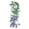 7qieC  2gk9S C: citing same article ( S: Starting model for refinement |
|---|---|
| Similar structure data |
-Links
 PDBj
PDBj


- Assembly
| Deposited unit | 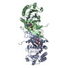
| ||||||||
|---|---|---|---|---|---|---|---|---|---|
| 1 | 
| ||||||||
| 2 | 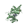
| ||||||||
| Unit cell |
|
-Components
| #1: Protein | Mass: 40148.898 Da / Num. of mol.: 2 / Mutation: aa32-421(delta 300-341) Source method: isolated from a genetically manipulated source Source: (gene. exp.) Homo sapiens (human) / Production host:  #2: Chemical | 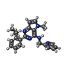  Mass: 357.452 Da / Num. of mol.: 2 / Source method: obtained synthetically / Formula: C22H23N5 / Feature type: SUBJECT OF INVESTIGATION Mass: 357.452 Da / Num. of mol.: 2 / Source method: obtained synthetically / Formula: C22H23N5 / Feature type: SUBJECT OF INVESTIGATION#3: Chemical |   Mass: 506.196 Da / Num. of mol.: 2 / Source method: obtained synthetically / Formula: C10H17N6O12P3 / Comment: AMP-PNP, energy-carrying molecule analogue*YM Mass: 506.196 Da / Num. of mol.: 2 / Source method: obtained synthetically / Formula: C10H17N6O12P3 / Comment: AMP-PNP, energy-carrying molecule analogue*YM#4: Water | ChemComp-HOH / |  Mass: 18.015 Da / Num. of mol.: 210 / Source method: isolated from a natural source / Formula: H2O Mass: 18.015 Da / Num. of mol.: 210 / Source method: isolated from a natural source / Formula: H2OHas ligand of interest | Y | |
|---|
-Experimental details
-Experiment
| Experiment | Method: X-RAY DIFFRACTION / Number of used crystals: 1 |
|---|
- Sample preparation
| Crystal | Density Matthews: 2.29 Å3/Da / Density % sol: 46.29 % |
|---|---|
| Crystal grow | Temperature: 293 K / Method: vapor diffusion, sitting drop Details: 20% Peg5KMME, 0.3M Ammonium Tartrate, 100mM PCTP, pH 7.5 |
-Data collection
| Diffraction | Mean temperature: 100 K / Serial crystal experiment: N |
|---|---|
| Diffraction source | Source: SYNCHROTRON / Site: Diamond / Beamline: I24 / Wavelength: 0.9999 Å |
| Detector | Type: DECTRIS EIGER2 X 9M / Detector: PIXEL / Date: Apr 30, 2021 |
| Radiation | Protocol: SINGLE WAVELENGTH / Monochromatic (M) / Laue (L): M / Scattering type: x-ray |
| Radiation wavelength | Wavelength: 0.9999 Å / Relative weight: 1 |
| Reflection | Resolution: 1.95→116.94 Å / Num. obs: 39543 / % possible obs: 91.7 % / Redundancy: 5.4 % / CC1/2: 0.996 / Net I/σ(I): 9.2 |
| Reflection shell | Resolution: 1.95→2.14 Å / Redundancy: 5.1 % / Num. unique obs: 1978 / CC1/2: 0.56 / % possible all: 56.5 |
- Processing
| Software |
| ||||||||||||||||||||||||||||||||||||||||||||||||||||||||||||
|---|---|---|---|---|---|---|---|---|---|---|---|---|---|---|---|---|---|---|---|---|---|---|---|---|---|---|---|---|---|---|---|---|---|---|---|---|---|---|---|---|---|---|---|---|---|---|---|---|---|---|---|---|---|---|---|---|---|---|---|---|---|
| Refinement | Method to determine structure: MOLECULAR REPLACEMENT Starting model: 2GK9 Resolution: 1.95→116.94 Å / Cor.coef. Fo:Fc: 0.958 / Cor.coef. Fo:Fc free: 0.919 / SU B: 6.015 / SU ML: 0.16 / Cross valid method: THROUGHOUT / σ(F): 0 / ESU R: 0.248 / ESU R Free: 0.218 / Stereochemistry target values: MAXIMUM LIKELIHOOD Details: HYDROGENS HAVE BEEN ADDED IN THE RIDING POSITIONS U VALUES : REFINED INDIVIDUALLY
| ||||||||||||||||||||||||||||||||||||||||||||||||||||||||||||
| Solvent computation | Ion probe radii: 0.8 Å / Shrinkage radii: 0.8 Å / VDW probe radii: 1.2 Å / Solvent model: MASK | ||||||||||||||||||||||||||||||||||||||||||||||||||||||||||||
| Displacement parameters | Biso max: 132.25 Å2 / Biso mean: 47.45 Å2 / Biso min: 10.19 Å2
| ||||||||||||||||||||||||||||||||||||||||||||||||||||||||||||
| Refinement step | Cycle: final / Resolution: 1.95→116.94 Å
| ||||||||||||||||||||||||||||||||||||||||||||||||||||||||||||
| Refine LS restraints |
| ||||||||||||||||||||||||||||||||||||||||||||||||||||||||||||
| LS refinement shell | Resolution: 1.95→1.997 Å / Rfactor Rfree error: 0
|
