 Movie
Movie Controller


+ Open data

- Basic information
| Entry | Database: PDB / ID: 7qp5 | ||||||||||||
|---|---|---|---|---|---|---|---|---|---|---|---|---|---|
| Title | Crystal Structure of E. coli FhuF | ||||||||||||
 Components Components | Ferric iron reductase protein FhuF | ||||||||||||
 Keywords Keywords | OXIDOREDUCTASE / Ferric Siderophore Reductase / Iron-Sulphur protein / Iron uptake | ||||||||||||
| Function / homology |  Function and homology information Function and homology informationferric-chelate reductase activity / reductive iron assimilation / siderophore-iron import into cell / oxidoreductase activity, acting on metal ions / 2 iron, 2 sulfur cluster binding / metal ion binding / plasma membrane / cytosol Similarity search - Function | ||||||||||||
| Biological species |  | ||||||||||||
| Method |  X-RAY DIFFRACTION / SYNCHROTRON / MOLECULAR REPLACEMENT / Resolution: 1.92 Å X-RAY DIFFRACTION / SYNCHROTRON / MOLECULAR REPLACEMENT / Resolution: 1.92 Å | ||||||||||||
 Authors Authors | Trindade, I.B. / Rollo, F. / Matias, P.M. / Moe, E. / Louro, R.O. | ||||||||||||
| Funding support |  Portugal, European Union, 3items Portugal, European Union, 3items
| ||||||||||||
 Citation Citation | Journal: Biorxiv / Year: 2023 Title: The structure of a novel ferredoxin: FhuF, a ferric-siderophore reductase from E. coli K-12 with a novel 2Fe-2S cluster coordination Authors: Trindade, I.B. / Rollo, F. / Todorovic, S. / Catarino, T. / Moe, E. / Matias, P.M. / Piccioli, M. / Louro, R.O. | ||||||||||||
| History |
|
- Structure visualization
| Structure viewer | Molecule: MolmilJmol/JSmol |
|---|
- Downloads & links
-Download
| PDBx/mmCIF format | 7qp5.cif.gz | 238 KB | Display | PDBx/mmCIF format |
|---|---|---|---|---|
| PDB format | pdb7qp5.ent.gz | 155.9 KB | Display | PDB format |
| PDBx/mmJSON format | 7qp5.json.gz | Tree view | PDBx/mmJSON format | |
| Others |  Other downloads Other downloads |
-Validation report
| Arichive directory | https://data.pdbj.org/pub/pdb/validation_reports/qp/7qp5ftp://data.pdbj.org/pub/pdb/validation_reports/qp/7qp5 | HTTPS FTP |
|---|
-Related structure data
| Similar structure data |
|---|
-Links
 PDBj
PDBj


- Assembly
| Deposited unit | 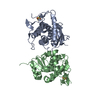
| |||||||||||||||||||||||||||||||||||||||||||||||||||||||||||
|---|---|---|---|---|---|---|---|---|---|---|---|---|---|---|---|---|---|---|---|---|---|---|---|---|---|---|---|---|---|---|---|---|---|---|---|---|---|---|---|---|---|---|---|---|---|---|---|---|---|---|---|---|---|---|---|---|---|---|---|---|
| 1 | 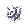
| |||||||||||||||||||||||||||||||||||||||||||||||||||||||||||
| 2 | 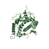
| |||||||||||||||||||||||||||||||||||||||||||||||||||||||||||
| Unit cell |
| |||||||||||||||||||||||||||||||||||||||||||||||||||||||||||
| Noncrystallographic symmetry (NCS) | NCS domain:
NCS domain segments: Ens-ID: ens_1
NCS oper: (Code: givenMatrix: (0.281609956578, -0.00462675931479, 0.959517808826), (0.000584849470999, -0.999987361124, -0.00499355012259), (0.959528785554, 0.0019674069161, -0.281603691387)Vector: - ...NCS oper: (Code: given Matrix: (0.281609956578, -0.00462675931479, 0.959517808826), Vector: |
-Components
| #1: Protein | Mass: 28037.258 Da / Num. of mol.: 2 Source method: isolated from a genetically manipulated source Source: (gene. exp.) #2: Chemical |   Mass: 175.820 Da / Num. of mol.: 2 / Source method: isolated from a natural source / Formula: Fe2S2 / Feature type: SUBJECT OF INVESTIGATION Mass: 175.820 Da / Num. of mol.: 2 / Source method: isolated from a natural source / Formula: Fe2S2 / Feature type: SUBJECT OF INVESTIGATION#3: Water | ChemComp-HOH / |  Mass: 18.015 Da / Num. of mol.: 157 / Source method: isolated from a natural source / Formula: H2O Mass: 18.015 Da / Num. of mol.: 157 / Source method: isolated from a natural source / Formula: H2OHas ligand of interest | Y | |
|---|
-Experimental details
-Experiment
| Experiment | Method: X-RAY DIFFRACTION / Number of used crystals: 1 |
|---|
- Sample preparation
| Crystal | Density Matthews: 2.55 Å3/Da / Density % sol: 51.78 % / Description: Thick plate |
|---|---|
| Crystal grow | Temperature: 298 K / Method: vapor diffusion, hanging drop / pH: 8.2 Details: Protein concentration 50 MG/ML, 0.2 M MGCL2.6H2O, 0.1 M TRIS-HCL PH 8.5, 30% W/V PEG 4000, Hanging drop vapor diffusion; 2 microliters protein + 2 microliters reservoir equilibrated against ...Details: Protein concentration 50 MG/ML, 0.2 M MGCL2.6H2O, 0.1 M TRIS-HCL PH 8.5, 30% W/V PEG 4000, Hanging drop vapor diffusion; 2 microliters protein + 2 microliters reservoir equilibrated against 500 microliters reservoir |
-Data collection
| Diffraction | Mean temperature: 100 K / Serial crystal experiment: N |
|---|---|
| Diffraction source | Source: SYNCHROTRON / Site: ALBA  / Beamline: XALOC / Wavelength: 0.97934 Å / Beamline: XALOC / Wavelength: 0.97934 Å |
| Detector | Type: DECTRIS PILATUS 6M / Detector: PIXEL / Date: Dec 5, 2021 |
| Radiation | Protocol: SINGLE WAVELENGTH / Monochromatic (M) / Laue (L): M / Scattering type: x-ray |
| Radiation wavelength | Wavelength: 0.97934 Å / Relative weight: 1 |
| Reflection | Resolution: 1.92→65.95 Å / Num. obs: 25252 / % possible obs: 52.7 % / Redundancy: 3.3 % / Biso Wilson estimate: 28.19 Å2 / CC1/2: 0.997 / Rmerge(I) obs: 0.078 / Rpim(I) all: 0.05 / Net I/σ(I): 9.3 |
| Reflection shell | Resolution: 1.92→2.11 Å / Redundancy: 2.1 % / Rmerge(I) obs: 0.567 / Mean I/σ(I) obs: 1.4 / Num. unique obs: 1127 / CC1/2: 0.56 / Rpim(I) all: 0.492 / % possible all: 10.7 |
- Processing
| Software |
| ||||||||||||||||||||||||||||||||||||||||||||||||||||||||||||||||||||||
|---|---|---|---|---|---|---|---|---|---|---|---|---|---|---|---|---|---|---|---|---|---|---|---|---|---|---|---|---|---|---|---|---|---|---|---|---|---|---|---|---|---|---|---|---|---|---|---|---|---|---|---|---|---|---|---|---|---|---|---|---|---|---|---|---|---|---|---|---|---|---|---|
| Refinement | Method to determine structure: MOLECULAR REPLACEMENT Starting model: ALPHA FOLD MODEL Resolution: 1.92→65.95 Å / SU ML: 0.2721 / Cross valid method: FREE R-VALUE / σ(F): 1.36 / Phase error: 32.1797 Stereochemistry target values: GeoStd + Monomer Library + CDL v1.2
| ||||||||||||||||||||||||||||||||||||||||||||||||||||||||||||||||||||||
| Solvent computation | Shrinkage radii: 0.9 Å / VDW probe radii: 1.11 Å / Solvent model: FLAT BULK SOLVENT MODEL | ||||||||||||||||||||||||||||||||||||||||||||||||||||||||||||||||||||||
| Displacement parameters | Biso mean: 33.81 Å2 | ||||||||||||||||||||||||||||||||||||||||||||||||||||||||||||||||||||||
| Refinement step | Cycle: LAST / Resolution: 1.92→65.95 Å
| ||||||||||||||||||||||||||||||||||||||||||||||||||||||||||||||||||||||
| Refine LS restraints |
| ||||||||||||||||||||||||||||||||||||||||||||||||||||||||||||||||||||||
| Refine LS restraints NCS | Type: Torsion NCS / Rms dev position: 0.213809848111 Å | ||||||||||||||||||||||||||||||||||||||||||||||||||||||||||||||||||||||
| LS refinement shell |
|
