 Movie
Movie Controller

[English] 日本語
 Yorodumi
- PDB-7qoa: Structure of CodB, a cytosine transporter in an outward-facing co... -
+ Open data

- Basic information
| Entry | Database: PDB / ID: 7qoa | ||||||
|---|---|---|---|---|---|---|---|
| Title | Structure of CodB, a cytosine transporter in an outward-facing conformation | ||||||
 Components Components | Cytosine permease | ||||||
 Keywords Keywords | TRANSPORT PROTEIN / Membrane Transporter / Cytosine / Sodium | ||||||
| Function / homology | cytosine transmembrane transporter activity / Cytosine permease / Purine-cytosine permease / Permease for cytosine/purines, uracil, thiamine, allantoin / membrane => GO:0016020 / 2,3-dihydroxypropyl (9Z)-octadec-9-enoate / 6-AMINOPYRIMIDIN-2(1H)-ONE / DI-PALMITOYL-3-SN-PHOSPHATIDYLETHANOLAMINE / Cytosine permease Function and homology information Function and homology information | ||||||
| Biological species |  Proteus vulgaris (bacteria) Proteus vulgaris (bacteria) | ||||||
| Method |  X-RAY DIFFRACTION / SYNCHROTRON / MOLECULAR REPLACEMENT / Resolution: 2.4 Å X-RAY DIFFRACTION / SYNCHROTRON / MOLECULAR REPLACEMENT / Resolution: 2.4 Å | ||||||
 Authors Authors | Hatton, C.E. / Cameron, A.D. | ||||||
| Funding support |  United Kingdom, 1items United Kingdom, 1items
| ||||||
 Citation Citation | Journal: Embo J. / Year: 2022 Title: Structure of cytosine transport protein CodB provides insight into nucleobase-cation symporter 1 mechanism. Authors: Hatton, C.E. / Brotherton, D.H. / Spencer, M. / Cameron, A.D. | ||||||
| History |
|
- Structure visualization
| Structure viewer | Molecule: MolmilJmol/JSmol |
|---|
- Downloads & links
-Download
| PDBx/mmCIF format | 7qoa.cif.gz | 200.6 KB | Display | PDBx/mmCIF format |
|---|---|---|---|---|
| PDB format | pdb7qoa.ent.gz | 125.9 KB | Display | PDB format |
| PDBx/mmJSON format | 7qoa.json.gz | Tree view | PDBx/mmJSON format | |
| Others |  Other downloads Other downloads |
-Validation report
| Summary document | 7qoa_validation.pdf.gz | 2.4 MB | Display | wwPDB validaton report |
|---|---|---|---|---|
| Full document | 7qoa_full_validation.pdf.gz | 2.4 MB | Display | |
| Data in XML | 7qoa_validation.xml.gz | 30.2 KB | Display | |
| Data in CIF | 7qoa_validation.cif.gz | 41.5 KB | Display | |
| Arichive directory | https://data.pdbj.org/pub/pdb/validation_reports/qo/7qoaftp://data.pdbj.org/pub/pdb/validation_reports/qo/7qoa | HTTPS FTP |
-Related structure data
| Related structure data | 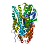 2jlnS S: Starting model for refinement |
|---|---|
| Similar structure data |
-Links
 PDBj
PDBj- Assembly
| Deposited unit | 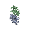
| ||||||||||||
|---|---|---|---|---|---|---|---|---|---|---|---|---|---|
| 1 | 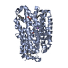
| ||||||||||||
| 2 | 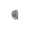
| ||||||||||||
| Unit cell |
|
-Components
-Protein / Sugars , 2 types, 3 molecules AB
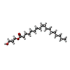
| #1: Protein | Mass: 44191.785 Da / Num. of mol.: 2 Source method: isolated from a genetically manipulated source Source: (gene. exp.) Proteus vulgaris (bacteria) / Gene: EKQ45_12260 / Production host: #4: Sugar | ChemComp-LMT / |  Type: D-saccharide / Mass: 510.615 Da / Num. of mol.: 1 / Source method: obtained synthetically / Formula: C24H46O11 / Comment: detergent*YM Type: D-saccharide / Mass: 510.615 Da / Num. of mol.: 1 / Source method: obtained synthetically / Formula: C24H46O11 / Comment: detergent*YM |
|---|
-Non-polymers , 5 types, 76 molecules 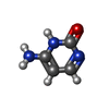


| #2: Chemical |  Mass: 111.102 Da / Num. of mol.: 2 / Source method: obtained synthetically / Formula: C4H5N3O / Feature type: SUBJECT OF INVESTIGATION Mass: 111.102 Da / Num. of mol.: 2 / Source method: obtained synthetically / Formula: C4H5N3O / Feature type: SUBJECT OF INVESTIGATION#3: Chemical | ChemComp-PEF / |  Mass: 691.959 Da / Num. of mol.: 1 / Source method: obtained synthetically / Formula: C37H74NO8P / Comment: phospholipid*YM Mass: 691.959 Da / Num. of mol.: 1 / Source method: obtained synthetically / Formula: C37H74NO8P / Comment: phospholipid*YM#5: Chemical | ChemComp-A6L /  Mass: 356.540 Da / Num. of mol.: 5 / Source method: obtained synthetically / Formula: C21H40O4 Mass: 356.540 Da / Num. of mol.: 5 / Source method: obtained synthetically / Formula: C21H40O4#6: Chemical |  Mass: 22.990 Da / Num. of mol.: 2 / Source method: isolated from a natural source / Formula: Na / Feature type: SUBJECT OF INVESTIGATION Mass: 22.990 Da / Num. of mol.: 2 / Source method: isolated from a natural source / Formula: Na / Feature type: SUBJECT OF INVESTIGATION#7: Water | ChemComp-HOH / | Mass: 18.015 Da / Num. of mol.: 66 / Source method: isolated from a natural source / Formula: H2O |
|---|
-Details
| Has ligand of interest | Y |
|---|
-Experimental details
-Experiment
| Experiment | Method: X-RAY DIFFRACTION / Number of used crystals: 1 |
|---|
- Sample preparation
| Crystal | Density Matthews: 3.28 Å3/Da / Density % sol: 62.48 % |
|---|---|
| Crystal grow | Temperature: 293 K / Method: lipidic cubic phase / pH: 6.5 Details: 0.1 M Sodium Cacodylate pH 6.5, 0.45 M NaCl, 39% PEG400 |
-Data collection
| Diffraction | Mean temperature: 100 K / Serial crystal experiment: N |
|---|---|
| Diffraction source | Source: SYNCHROTRON / Site: Diamond / Beamline: I24 / Wavelength: 0.9686 Å |
| Detector | Type: DECTRIS PILATUS3 6M / Detector: PIXEL / Date: Oct 11, 2019 |
| Radiation | Protocol: SINGLE WAVELENGTH / Monochromatic (M) / Laue (L): M / Scattering type: x-ray |
| Radiation wavelength | Wavelength: 0.9686 Å / Relative weight: 1 |
| Reflection | Resolution: 2.4→73.16 Å / Num. obs: 45754 / % possible obs: 99.41 % / Redundancy: 12.7 % / Biso Wilson estimate: 42.05 Å2 / CC1/2: 0.998 / Rmerge(I) obs: 0.3739 / Net I/σ(I): 11.57 |
| Reflection shell | Resolution: 2.4→2.49 Å / Num. unique obs: 4436 / CC1/2: 0.466 |
- Processing
| Software |
| |||||||||||||||||||||||||||||||||||||||||||||||||||||||||||||||||||||||||||||||||||||||||||||||||||||||||||||||||||||||
|---|---|---|---|---|---|---|---|---|---|---|---|---|---|---|---|---|---|---|---|---|---|---|---|---|---|---|---|---|---|---|---|---|---|---|---|---|---|---|---|---|---|---|---|---|---|---|---|---|---|---|---|---|---|---|---|---|---|---|---|---|---|---|---|---|---|---|---|---|---|---|---|---|---|---|---|---|---|---|---|---|---|---|---|---|---|---|---|---|---|---|---|---|---|---|---|---|---|---|---|---|---|---|---|---|---|---|---|---|---|---|---|---|---|---|---|---|---|---|---|---|
| Refinement | Method to determine structure: MOLECULAR REPLACEMENT Starting model: 2JLN Resolution: 2.4→73.16 Å / SU ML: 0.331 / Cross valid method: FREE R-VALUE / σ(F): 1.34 / Phase error: 28.1704 Stereochemistry target values: GeoStd + Monomer Library + CDL v1.2
| |||||||||||||||||||||||||||||||||||||||||||||||||||||||||||||||||||||||||||||||||||||||||||||||||||||||||||||||||||||||
| Solvent computation | Shrinkage radii: 0.9 Å / VDW probe radii: 1.11 Å / Solvent model: FLAT BULK SOLVENT MODEL | |||||||||||||||||||||||||||||||||||||||||||||||||||||||||||||||||||||||||||||||||||||||||||||||||||||||||||||||||||||||
| Displacement parameters | Biso mean: 47.52 Å2 | |||||||||||||||||||||||||||||||||||||||||||||||||||||||||||||||||||||||||||||||||||||||||||||||||||||||||||||||||||||||
| Refinement step | Cycle: LAST / Resolution: 2.4→73.16 Å
| |||||||||||||||||||||||||||||||||||||||||||||||||||||||||||||||||||||||||||||||||||||||||||||||||||||||||||||||||||||||
| Refine LS restraints |
| |||||||||||||||||||||||||||||||||||||||||||||||||||||||||||||||||||||||||||||||||||||||||||||||||||||||||||||||||||||||
| LS refinement shell |
|
