 Movie
Movie Controller

[English] 日本語
 Yorodumi
- PDB-7qo8: Structure of Protease1 from Pyrococcus horikoshii in space group ... -
+ Open data

- Basic information
| Entry | Database: PDB / ID: 7qo8 | ||||||
|---|---|---|---|---|---|---|---|
| Title | Structure of Protease1 from Pyrococcus horikoshii in space group 19 with a hexamer in the asymmetric unit | ||||||
 Components Components | Deglycase PH1704 | ||||||
 Keywords Keywords | HYDROLASE / protease 1 / hexamer | ||||||
| Function / homology |  Function and homology information Function and homology informationprotein deglycase / protein deglycase activity / peptidase activity / Hydrolases; Acting on peptide bonds (peptidases); Cysteine endopeptidases / proteolysis / cytoplasm Similarity search - Function | ||||||
| Biological species |   Pyrococcus horikoshii (archaea) Pyrococcus horikoshii (archaea) | ||||||
| Method |  X-RAY DIFFRACTION / SYNCHROTRON / MOLECULAR REPLACEMENT / Resolution: 1.95 Å X-RAY DIFFRACTION / SYNCHROTRON / MOLECULAR REPLACEMENT / Resolution: 1.95 Å | ||||||
 Authors Authors | Engilberge, S. / Gabel, F. / Girard, E. | ||||||
| Funding support |  France, 1items France, 1items
| ||||||
 Citation Citation | Journal: Acta Crystallogr D Struct Biol / Year: 2022 Title: Medical contrast agents as promising tools for biomacromolecular SAXS experiments. Authors: Gabel, F. / Engilberge, S. / Schmitt, E. / Thureau, A. / Mechulam, Y. / Perez, J. / Girard, E. | ||||||
| History |
|
- Structure visualization
| Structure viewer | Molecule: MolmilJmol/JSmol |
|---|
- Downloads & links
-Download
| PDBx/mmCIF format | 7qo8.cif.gz | 246.9 KB | Display | PDBx/mmCIF format |
|---|---|---|---|---|
| PDB format | pdb7qo8.ent.gz | 180.7 KB | Display | PDB format |
| PDBx/mmJSON format | 7qo8.json.gz | Tree view | PDBx/mmJSON format | |
| Others |  Other downloads Other downloads |
-Validation report
| Arichive directory | https://data.pdbj.org/pub/pdb/validation_reports/qo/7qo8ftp://data.pdbj.org/pub/pdb/validation_reports/qo/7qo8 | HTTPS FTP |
|---|
-Related structure data
| Related structure data | 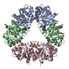 6q3tS S: Starting model for refinement |
|---|---|
| Similar structure data |
-Links
 PDBj
PDBj

- Assembly
| Deposited unit | 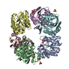
| |||||||||||||||||||||||||||||||||||||||||||||||||||||||||||||||||||||||||||||||||||||||||||||||
|---|---|---|---|---|---|---|---|---|---|---|---|---|---|---|---|---|---|---|---|---|---|---|---|---|---|---|---|---|---|---|---|---|---|---|---|---|---|---|---|---|---|---|---|---|---|---|---|---|---|---|---|---|---|---|---|---|---|---|---|---|---|---|---|---|---|---|---|---|---|---|---|---|---|---|---|---|---|---|---|---|---|---|---|---|---|---|---|---|---|---|---|---|---|---|---|---|
| 1 |
| |||||||||||||||||||||||||||||||||||||||||||||||||||||||||||||||||||||||||||||||||||||||||||||||
| Unit cell |
| |||||||||||||||||||||||||||||||||||||||||||||||||||||||||||||||||||||||||||||||||||||||||||||||
| Noncrystallographic symmetry (NCS) | NCS domain:
NCS domain segments: Ens-ID: 1
|