 Movie
Movie Controller

[English] 日本語
 Yorodumi
- PDB-7qh3: Crystal structure of the anti-sigma factor RsfG from Streptomyces... -
+ Open data

- Basic information
| Entry | Database: PDB / ID: 7qh3 | ||||||
|---|---|---|---|---|---|---|---|
| Title | Crystal structure of the anti-sigma factor RsfG from Streptomyces tsukubaensis NRRL18488 | ||||||
 Components Components | RsfG | ||||||
 Keywords Keywords | TRANSCRIPTION / anti-sigma / Streptomyces / sigma | ||||||
| Function / homology | DUF1579 domain-containing protein Function and homology information Function and homology information | ||||||
| Biological species |  Streptomyces tsukubensis NRRL18488 (bacteria) Streptomyces tsukubensis NRRL18488 (bacteria) | ||||||
| Method |  X-RAY DIFFRACTION / SYNCHROTRON / MOLECULAR REPLACEMENT / Resolution: 2.3 Å X-RAY DIFFRACTION / SYNCHROTRON / MOLECULAR REPLACEMENT / Resolution: 2.3 Å | ||||||
 Authors Authors | Leite, J.P. / Lourenco, F. / Gales, L. | ||||||
| Funding support |  Portugal, 1items Portugal, 1items
| ||||||
 Citation Citation | Journal: J.Struct.Biol. / Year: 2023 Title: Crystal structures of Streptomyces tsukubaensis sigma factor SigG1 and anti-sigma RsfG. Authors: Leite, J.P. / Lourenco, F. / Oliveira, R. / Sousa, S.F. / Mendes, M.V. / Gales, L. | ||||||
| History |
|
- Structure visualization
| Structure viewer | Molecule: MolmilJmol/JSmol |
|---|
- Downloads & links
-Download
| PDBx/mmCIF format | 7qh3.cif.gz | 119.2 KB | Display | PDBx/mmCIF format |
|---|---|---|---|---|
| PDB format | pdb7qh3.ent.gz | 91.8 KB | Display | PDB format |
| PDBx/mmJSON format | 7qh3.json.gz | Tree view | PDBx/mmJSON format | |
| Others |  Other downloads Other downloads |
-Validation report
| Summary document | 7qh3_validation.pdf.gz | 436.9 KB | Display | wwPDB validaton report |
|---|---|---|---|---|
| Full document | 7qh3_full_validation.pdf.gz | 439.2 KB | Display | |
| Data in XML | 7qh3_validation.xml.gz | 21.6 KB | Display | |
| Data in CIF | 7qh3_validation.cif.gz | 30.2 KB | Display | |
| Arichive directory | https://data.pdbj.org/pub/pdb/validation_reports/qh/7qh3ftp://data.pdbj.org/pub/pdb/validation_reports/qh/7qh3 | HTTPS FTP |
-Related structure data

-Links
 PDBj
PDBj- Assembly
| Deposited unit | 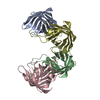
| ||||||||
|---|---|---|---|---|---|---|---|---|---|
| 1 | 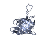
| ||||||||
| 2 | 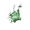
| ||||||||
| 3 | 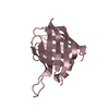
| ||||||||
| 4 | 
| ||||||||
| Unit cell |
|
-Components
| #1: Protein | Mass: 16351.823 Da / Num. of mol.: 4 Source method: isolated from a genetically manipulated source Source: (gene. exp.) Streptomyces tsukubensis NRRL18488 (bacteria)Strain: DSM 42081 / NBRC 108919 / NRRL 18488 / 9993 / Gene: STSU_011565, STSU_11555 / Production host: #2: Water | ChemComp-HOH / |  Mass: 18.015 Da / Num. of mol.: 139 / Source method: isolated from a natural source / Formula: H2O Mass: 18.015 Da / Num. of mol.: 139 / Source method: isolated from a natural source / Formula: H2O |
|---|
-Experimental details
-Experiment
| Experiment | Method: X-RAY DIFFRACTION / Number of used crystals: 1 |
|---|
- Sample preparation
| Crystal | Density Matthews: 2.72 Å3/Da / Density % sol: 54.73 % |
|---|---|
| Crystal grow | Temperature: 293 K / Method: vapor diffusion, sitting drop / pH: 8.5 Details: 0.1 M Ammonium phosphate dibasic; 0.1 M Tris hydrochloride pH 8.5; 0.5 M sodium phosphate dibasic dehydrate; 0.5 M potassium phosphate dibasic |
-Data collection
| Diffraction | Mean temperature: 100 K / Serial crystal experiment: N |
|---|---|
| Diffraction source | Source: SYNCHROTRON / Site: SOLEIL  / Beamline: PROXIMA 1 / Wavelength: 0.97856 Å / Beamline: PROXIMA 1 / Wavelength: 0.97856 Å |
| Detector | Type: DECTRIS PILATUS 6M / Detector: PIXEL / Date: May 25, 2018 |
| Radiation | Protocol: SINGLE WAVELENGTH / Monochromatic (M) / Laue (L): M / Scattering type: x-ray |
| Radiation wavelength | Wavelength: 0.97856 Å / Relative weight: 1 |
| Reflection | Resolution: 2.3→70.27 Å / Num. obs: 31236 / % possible obs: 99.5 % / Redundancy: 3.5 % / Biso Wilson estimate: 43.61 Å2 / CC1/2: 0.995 / Net I/σ(I): 8.5 |
| Reflection shell | Resolution: 2.3→2.38 Å / Rmerge(I) obs: 0.087 / Num. unique obs: 3091 |
- Processing
| Software |
| ||||||||||||||||||||||||||||||||||||||||||||||||||||||||||||||||||||||||||||||||||||
|---|---|---|---|---|---|---|---|---|---|---|---|---|---|---|---|---|---|---|---|---|---|---|---|---|---|---|---|---|---|---|---|---|---|---|---|---|---|---|---|---|---|---|---|---|---|---|---|---|---|---|---|---|---|---|---|---|---|---|---|---|---|---|---|---|---|---|---|---|---|---|---|---|---|---|---|---|---|---|---|---|---|---|---|---|---|
| Refinement | Method to determine structure: MOLECULAR REPLACEMENT / Resolution: 2.3→70.27 Å / SU ML: 0.3 / Cross valid method: THROUGHOUT / σ(F): 1.35 / Phase error: 23.82 / Stereochemistry target values: ML
| ||||||||||||||||||||||||||||||||||||||||||||||||||||||||||||||||||||||||||||||||||||
| Solvent computation | Shrinkage radii: 0.9 Å / VDW probe radii: 1.11 Å / Solvent model: FLAT BULK SOLVENT MODEL | ||||||||||||||||||||||||||||||||||||||||||||||||||||||||||||||||||||||||||||||||||||
| Displacement parameters | Biso max: 95.75 Å2 / Biso mean: 44.9133 Å2 / Biso min: 22.19 Å2 | ||||||||||||||||||||||||||||||||||||||||||||||||||||||||||||||||||||||||||||||||||||
| Refinement step | Cycle: final / Resolution: 2.3→70.27 Å
| ||||||||||||||||||||||||||||||||||||||||||||||||||||||||||||||||||||||||||||||||||||
| LS refinement shell | Refine-ID: X-RAY DIFFRACTION / Rfactor Rfree error: 0 / Total num. of bins used: 11
|
