 Movie
Movie Controller

[English] 日本語
 Yorodumi
- PDB-7qf6: N(5)-hydroxyornithine:cis-anhydromevalonyl coenzyme A-N(5)-transa... -
+ Open data

- Basic information
| Entry | Database: PDB / ID: 7qf6 | ||||||
|---|---|---|---|---|---|---|---|
| Title | N(5)-hydroxyornithine:cis-anhydromevalonyl coenzyme A-N(5)-transacylase sidF | ||||||
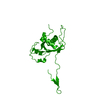
 Components Components | N(5)-hydroxyornithine:cis-anhydromevalonyl coenzyme A-N(5)-transacylase sidF | ||||||
 Keywords Keywords | TRANSFERASE / Hydroxyornithine transacylase | ||||||
| Function / homology |  Function and homology information Function and homology informationN',N'',N'''-triacetylfusarinine C biosynthetic process / ergosterol biosynthetic process / : / siderophore biosynthetic process / Transferases; Acyltransferases; Transferring groups other than aminoacyl groups / peroxisome Similarity search - Function | ||||||
| Biological species |  | ||||||
| Method |  X-RAY DIFFRACTION / SYNCHROTRON / MOLECULAR REPLACEMENT / Resolution: 1.87 Å X-RAY DIFFRACTION / SYNCHROTRON / MOLECULAR REPLACEMENT / Resolution: 1.87 Å | ||||||
 Authors Authors | Poonsiri, T. / Demitri, N. / Stefano, B. | ||||||
| Funding support | European Union, 1items
| ||||||
 Citation Citation | Journal: J Struct Biol X / Year: 2025 Title: SidF, a dual substrate N5-acetyl-N5-hydroxy-L-ornithine transacetylase involved in Aspergillus fumigatus siderophore biosynthesis. Authors: Poonsiri, T. / Stransky, J. / Demitri, N. / Haas, H. / Cianci, M. / Benini, S. | ||||||
| History |
|
- Structure visualization
| Structure viewer | Molecule: MolmilJmol/JSmol |
|---|
- Downloads & links
-Download
| PDBx/mmCIF format | 7qf6.cif.gz | 746.8 KB | Display | PDBx/mmCIF format |
|---|---|---|---|---|
| PDB format | pdb7qf6.ent.gz | 613.7 KB | Display | PDB format |
| PDBx/mmJSON format | 7qf6.json.gz | Tree view | PDBx/mmJSON format | |
| Others |  Other downloads Other downloads |
-Validation report
| Arichive directory | https://data.pdbj.org/pub/pdb/validation_reports/qf/7qf6ftp://data.pdbj.org/pub/pdb/validation_reports/qf/7qf6 | HTTPS FTP |
|---|
-Related structure data
-Links
 PDBj
PDBj


- Assembly
| Deposited unit | 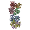
| ||||||||
|---|---|---|---|---|---|---|---|---|---|
| 1 | 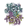
| ||||||||
| 2 | 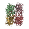
| ||||||||
| Unit cell |
|
-Components
| #1: Protein | Mass: 53377.723 Da / Num. of mol.: 8 Source method: isolated from a genetically manipulated source Source: (gene. exp.)  References: UniProt: Q4WF55, Transferases; Acyltransferases; Transferring groups other than aminoacyl groups #2: Chemical | ChemComp-GOL /   Mass: 92.094 Da / Num. of mol.: 17 / Source method: obtained synthetically / Formula: C3H8O3 Mass: 92.094 Da / Num. of mol.: 17 / Source method: obtained synthetically / Formula: C3H8O3#3: Chemical | ChemComp-SCN /   Mass: 58.082 Da / Num. of mol.: 4 / Source method: obtained synthetically / Formula: CNS Mass: 58.082 Da / Num. of mol.: 4 / Source method: obtained synthetically / Formula: CNS#4: Chemical |   Mass: 39.098 Da / Num. of mol.: 2 / Source method: obtained synthetically / Formula: K Mass: 39.098 Da / Num. of mol.: 2 / Source method: obtained synthetically / Formula: K#5: Water | ChemComp-HOH / |  Mass: 18.015 Da / Num. of mol.: 1973 / Source method: isolated from a natural source / Formula: H2O Mass: 18.015 Da / Num. of mol.: 1973 / Source method: isolated from a natural source / Formula: H2OHas ligand of interest | N | Has protein modification | N | |
|---|
-Experimental details
-Experiment
| Experiment | Method: X-RAY DIFFRACTION / Number of used crystals: 1 |
|---|
- Sample preparation
| Crystal | Density Matthews: 2.27 Å3/Da / Density % sol: 45.74 % |
|---|---|
| Crystal grow | Temperature: 293 K / Method: vapor diffusion, sitting drop / Details: 18-20% PEG 3350, 0.2-0.24 M potassium thiocyanate |
-Data collection
| Diffraction | Mean temperature: 80 K / Serial crystal experiment: N | ||||||||||||||||||||||||||||||
|---|---|---|---|---|---|---|---|---|---|---|---|---|---|---|---|---|---|---|---|---|---|---|---|---|---|---|---|---|---|---|---|
| Diffraction source | Source: SYNCHROTRON / Site: ELETTRA  / Beamline: 11.2C / Wavelength: 1 Å / Beamline: 11.2C / Wavelength: 1 Å | ||||||||||||||||||||||||||||||
| Detector | Type: DECTRIS PILATUS 6M / Detector: PIXEL / Date: Sep 28, 2021 | ||||||||||||||||||||||||||||||
| Radiation | Monochromator: Si(111) DCM / Protocol: SINGLE WAVELENGTH / Monochromatic (M) / Laue (L): M / Scattering type: x-ray | ||||||||||||||||||||||||||||||
| Radiation wavelength | Wavelength: 1 Å / Relative weight: 1 | ||||||||||||||||||||||||||||||
| Reflection | Resolution: 1.87→174.82 Å / Num. obs: 313786 / % possible obs: 97.1 % / Redundancy: 6.8 % / CC1/2: 0.999 / Rmerge(I) obs: 0.066 / Rpim(I) all: 0.027 / Rrim(I) all: 0.071 / Net I/σ(I): 17 / Num. measured all: 2129682 / Scaling rejects: 12 | ||||||||||||||||||||||||||||||
| Reflection shell | Diffraction-ID: 1
|
- Processing
| Software |
| |||||||||||||||||||||||||||||||||||||||||||||
|---|---|---|---|---|---|---|---|---|---|---|---|---|---|---|---|---|---|---|---|---|---|---|---|---|---|---|---|---|---|---|---|---|---|---|---|---|---|---|---|---|---|---|---|---|---|---|
| Refinement | Method to determine structure: MOLECULAR REPLACEMENT Starting model: AlphaFold Resolution: 1.87→174.82 Å / Cor.coef. Fo:Fc: 0.966 / Cor.coef. Fo:Fc free: 0.95 / SU B: 3.189 / SU ML: 0.093 / SU R Cruickshank DPI: 0.139 / Cross valid method: THROUGHOUT / σ(F): 0 / ESU R: 0.139 / ESU R Free: 0.129 / Stereochemistry target values: MAXIMUM LIKELIHOOD Details: HYDROGENS HAVE BEEN USED IF PRESENT IN THE INPUT U VALUES : REFINED INDIVIDUALLY
| |||||||||||||||||||||||||||||||||||||||||||||
| Solvent computation | Ion probe radii: 0.8 Å / Shrinkage radii: 0.8 Å / VDW probe radii: 1.2 Å / Solvent model: MASK | |||||||||||||||||||||||||||||||||||||||||||||
| Displacement parameters | Biso max: 124.06 Å2 / Biso mean: 33.442 Å2 / Biso min: 14.52 Å2
| |||||||||||||||||||||||||||||||||||||||||||||
| Refinement step | Cycle: final / Resolution: 1.87→174.82 Å
| |||||||||||||||||||||||||||||||||||||||||||||
| Refine LS restraints |
| |||||||||||||||||||||||||||||||||||||||||||||
| LS refinement shell | Resolution: 1.87→1.919 Å / Rfactor Rfree error: 0 / Total num. of bins used: 20
|
