 Movie
Movie Controller

[English] 日本語
 Yorodumi
- PDB-7qd0: Structure of the orange carotenoid protein from Planktothrix agar... -
+ Open data

- Basic information
| Entry | Database: PDB / ID: 7qd0 | |||||||||||||||
|---|---|---|---|---|---|---|---|---|---|---|---|---|---|---|---|---|
| Title | Structure of the orange carotenoid protein from Planktothrix agardhii binding echinenone in the C2 space group | |||||||||||||||
 Components Components | Orange carotenoid-binding protein | |||||||||||||||
 Keywords Keywords | PHOTOSYNTHESIS / CAROTENOID-BINDING / PHOTOPROTECTION / CAROTENOID BINDING PROTEIN | |||||||||||||||
| Function / homology |  Function and homology information Function and homology information | |||||||||||||||
| Biological species |  Planktothrix agardhii (bacteria) Planktothrix agardhii (bacteria) | |||||||||||||||
| Method |  X-RAY DIFFRACTION / SYNCHROTRON / MOLECULAR REPLACEMENT / Resolution: 1.7 Å X-RAY DIFFRACTION / SYNCHROTRON / MOLECULAR REPLACEMENT / Resolution: 1.7 Å | |||||||||||||||
 Authors Authors | Andreeva, E.A. / Hartmann, E. / Schlichting, I. / Colletier, J.-P. | |||||||||||||||
| Funding support |  France, 4items France, 4items
| |||||||||||||||
 Citation Citation | Journal: Biochim Biophys Acta Bioenerg / Year: 2022 Title: Structure-function-dynamics relationships in the peculiar Planktothrix PCC7805 OCP1: Impact of his-tagging and carotenoid type. Authors: Wilson, A. / Andreeva, E.A. / Nizinski, S.J. / Talbot, L. / Hartmann, E. / Schlichting, I. / Burdzinski, G. / Sliwa, M. / Kirilovsky, D. / Colletier, J.P. | |||||||||||||||
| History |
|
- Structure visualization
| Structure viewer | Molecule: MolmilJmol/JSmol |
|---|
- Downloads & links
-Download
| PDBx/mmCIF format | 7qd0.cif.gz | 107.1 KB | Display | PDBx/mmCIF format |
|---|---|---|---|---|
| PDB format | pdb7qd0.ent.gz | 66.3 KB | Display | PDB format |
| PDBx/mmJSON format | 7qd0.json.gz | Tree view | PDBx/mmJSON format | |
| Others |  Other downloads Other downloads |
-Validation report
| Arichive directory | https://data.pdbj.org/pub/pdb/validation_reports/qd/7qd0ftp://data.pdbj.org/pub/pdb/validation_reports/qd/7qd0 | HTTPS FTP |
|---|
-Related structure data
| Related structure data | 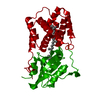 7qczC  7qd1C 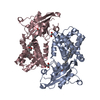 7qd2C  3mg1S S: Starting model for refinement C: citing same article ( |
|---|---|
| Similar structure data |
-Links
 PDBj
PDBj

- Assembly
| Deposited unit | 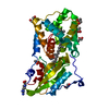
| |||||||||||||||
|---|---|---|---|---|---|---|---|---|---|---|---|---|---|---|---|---|
| 1 | 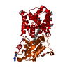
| |||||||||||||||
| Unit cell |
| |||||||||||||||
| Components on special symmetry positions |
|
-Components
-Protein , 1 types, 1 molecules A
| #1: Protein | Mass: 35352.434 Da / Num. of mol.: 1 Source method: isolated from a genetically manipulated source Source: (gene. exp.) Planktothrix agardhii (bacteria) / Gene: PLAM_2315Production host: References: UniProt: A0A1J1JHR9 |
|---|
-Non-polymers , 5 types, 268 molecules 


| #2: Chemical | ChemComp-ARG /  Type: L-peptide linking / Mass: 175.209 Da / Num. of mol.: 1 / Source method: obtained synthetically / Formula: C6H15N4O2 Type: L-peptide linking / Mass: 175.209 Da / Num. of mol.: 1 / Source method: obtained synthetically / Formula: C6H15N4O2 | ||||||
|---|---|---|---|---|---|---|---|
| #3: Chemical | ChemComp-GOL /  Mass: 92.094 Da / Num. of mol.: 5 / Source method: obtained synthetically / Formula: C3H8O3 Mass: 92.094 Da / Num. of mol.: 5 / Source method: obtained synthetically / Formula: C3H8O3#4: Chemical |  Mass: 59.044 Da / Num. of mol.: 2 / Source method: obtained synthetically / Formula: C2H3O2 Mass: 59.044 Da / Num. of mol.: 2 / Source method: obtained synthetically / Formula: C2H3O2#5: Chemical | ChemComp-ECH / |  Mass: 550.856 Da / Num. of mol.: 1 / Source method: obtained synthetically / Formula: C40H54O / Feature type: SUBJECT OF INVESTIGATION Mass: 550.856 Da / Num. of mol.: 1 / Source method: obtained synthetically / Formula: C40H54O / Feature type: SUBJECT OF INVESTIGATION#6: Water | ChemComp-HOH / | Mass: 18.015 Da / Num. of mol.: 259 / Source method: isolated from a natural source / Formula: H2O |
-Details
| Has ligand of interest | Y |
|---|
-Experimental details
-Experiment
| Experiment | Method: X-RAY DIFFRACTION / Number of used crystals: 1 |
|---|
- Sample preparation
| Crystal | Density Matthews: 2.12 Å3/Da / Density % sol: 41.93 % |
|---|---|
| Crystal grow | Temperature: 293 K / Method: vapor diffusion, hanging drop / pH: 5 / Details: 0.1 M sodium acetate, pH 5 and 10-20% PEG4000 |
-Data collection
| Diffraction | Mean temperature: 100 K / Serial crystal experiment: N |
|---|---|
| Diffraction source | Source: SYNCHROTRON / Site: ESRF / Beamline: ID29 / Wavelength: 0.9537 Å |
| Detector | Type: DECTRIS PILATUS 6M / Detector: PIXEL / Date: Jul 2, 2018 |
| Radiation | Protocol: SINGLE WAVELENGTH / Monochromatic (M) / Laue (L): M / Scattering type: x-ray |
| Radiation wavelength | Wavelength: 0.9537 Å / Relative weight: 1 |
| Reflection | Resolution: 1.7→32.44 Å / Num. obs: 30918 / % possible obs: 98.61 % / Redundancy: 3.3 % / Biso Wilson estimate: 25.58 Å2 / CC1/2: 0.999 / Rmerge(I) obs: 0.046 / Rpim(I) all: 0.03 / Rrim(I) all: 0.055 / Net I/σ(I): 13.78 |
| Reflection shell | Resolution: 1.7→1.76 Å / Redundancy: 3.4 % / Rmerge(I) obs: 0.7233 / Mean I/σ(I) obs: 1.85 / Num. unique obs: 3107 / CC1/2: 0.843 / Rpim(I) all: 0.3882 / Rrim(I) all: 0.7233 / % possible all: 99.42 |
- Processing
| Software |
| ||||||||||||||||||||||||||||||||||||||||||||||||||||||||||||||||||||||||||||||||||||
|---|---|---|---|---|---|---|---|---|---|---|---|---|---|---|---|---|---|---|---|---|---|---|---|---|---|---|---|---|---|---|---|---|---|---|---|---|---|---|---|---|---|---|---|---|---|---|---|---|---|---|---|---|---|---|---|---|---|---|---|---|---|---|---|---|---|---|---|---|---|---|---|---|---|---|---|---|---|---|---|---|---|---|---|---|---|
| Refinement | Method to determine structure: MOLECULAR REPLACEMENT Starting model: 3mg1 Resolution: 1.7→32.44 Å / SU ML: 0.2059 / Cross valid method: FREE R-VALUE / σ(F): 1.35 / Phase error: 26.2509 Stereochemistry target values: GeoStd + Monomer Library + CDL v1.2
| ||||||||||||||||||||||||||||||||||||||||||||||||||||||||||||||||||||||||||||||||||||
| Solvent computation | Shrinkage radii: 0.9 Å / VDW probe radii: 1.11 Å / Solvent model: FLAT BULK SOLVENT MODEL | ||||||||||||||||||||||||||||||||||||||||||||||||||||||||||||||||||||||||||||||||||||
| Displacement parameters | Biso mean: 34.32 Å2 | ||||||||||||||||||||||||||||||||||||||||||||||||||||||||||||||||||||||||||||||||||||
| Refinement step | Cycle: LAST / Resolution: 1.7→32.44 Å
| ||||||||||||||||||||||||||||||||||||||||||||||||||||||||||||||||||||||||||||||||||||
| Refine LS restraints |
| ||||||||||||||||||||||||||||||||||||||||||||||||||||||||||||||||||||||||||||||||||||
| LS refinement shell |
|
