 Movie
Movie Controller

[English] 日本語
 Yorodumi
- PDB-7qcf: X-ray crystallographic structure of E. coli K-12 glycyl-tRNA synt... -
+ Open data

- Basic information
| Entry | Database: PDB / ID: 7qcf | ||||||
|---|---|---|---|---|---|---|---|
| Title | X-ray crystallographic structure of E. coli K-12 glycyl-tRNA synthetase alpha subunit (glyQ) | ||||||
 Components Components | Glycine--tRNA ligase alpha subunit | ||||||
 Keywords Keywords | LIGASE / tRNA glycine | ||||||
| Function / homology |  Function and homology information Function and homology informationglycine-tRNA ligase complex / glycyl-tRNA aminoacylation / glycine-tRNA ligase / glycine-tRNA ligase activity / ATP binding / cytosol Similarity search - Function | ||||||
| Biological species |  | ||||||
| Method |  X-RAY DIFFRACTION / SYNCHROTRON / MOLECULAR REPLACEMENT / Resolution: 3 Å X-RAY DIFFRACTION / SYNCHROTRON / MOLECULAR REPLACEMENT / Resolution: 3 Å | ||||||
 Authors Authors | Weeks, S.D. / Munawar, A.H. | ||||||
| Funding support | 1items
| ||||||
 Citation Citation | Journal: To Be Published Title: Structure of E. coli K-12 glycyl-tRNA synthetase alpha subunit (glyQ) Authors: Weeks, S.D. / Munawar, A.H. | ||||||
| History |
|
- Structure visualization
| Structure viewer | Molecule: MolmilJmol/JSmol |
|---|
- Downloads & links
-Download
| PDBx/mmCIF format | 7qcf.cif.gz | 231.2 KB | Display | PDBx/mmCIF format |
|---|---|---|---|---|
| PDB format | pdb7qcf.ent.gz | 187 KB | Display | PDB format |
| PDBx/mmJSON format | 7qcf.json.gz | Tree view | PDBx/mmJSON format | |
| Others |  Other downloads Other downloads |
-Validation report
| Summary document | 7qcf_validation.pdf.gz | 456.5 KB | Display | wwPDB validaton report |
|---|---|---|---|---|
| Full document | 7qcf_full_validation.pdf.gz | 482.4 KB | Display | |
| Data in XML | 7qcf_validation.xml.gz | 41.4 KB | Display | |
| Data in CIF | 7qcf_validation.cif.gz | 55.7 KB | Display | |
| Arichive directory | https://data.pdbj.org/pub/pdb/validation_reports/qc/7qcfftp://data.pdbj.org/pub/pdb/validation_reports/qc/7qcf | HTTPS FTP |
-Related structure data
| Related structure data |  5f5wS S: Starting model for refinement |
|---|---|
| Similar structure data |
-Links
 PDBj
PDBj
- Assembly
| Deposited unit | 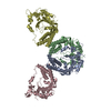
| ||||||||
|---|---|---|---|---|---|---|---|---|---|
| 1 | 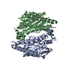
| ||||||||
| 2 | 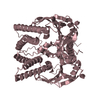
| ||||||||
| 3 | 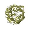
| ||||||||
| Unit cell |
|
-Components
| #1: Protein | Mass: 34876.242 Da / Num. of mol.: 4 Source method: isolated from a genetically manipulated source Source: (gene. exp.) Strain: K12 / Gene: glyQ, glyS(A), b3560, JW3531 / Plasmid: pET28 / Production host: |
|---|
-Experimental details
-Experiment
| Experiment | Method: X-RAY DIFFRACTION / Number of used crystals: 1 |
|---|
- Sample preparation
| Crystal | Density Matthews: 5.98 Å3/Da / Density % sol: 79.43 % |
|---|---|
| Crystal grow | Temperature: 293 K / Method: vapor diffusion, hanging drop / pH: 6.5 Details: 100 mM Morpheus buffer system 1, 0.5-3% w/v PEG 8000 and 20 % w/v ethylene glycol |
-Data collection
| Diffraction | Mean temperature: 100 K / Serial crystal experiment: N | ||||||||||||||||||||||||||||||
|---|---|---|---|---|---|---|---|---|---|---|---|---|---|---|---|---|---|---|---|---|---|---|---|---|---|---|---|---|---|---|---|
| Diffraction source | Source: SYNCHROTRON / Site: CHESS  / Beamline: A1 / Wavelength: 0.9775 Å / Beamline: A1 / Wavelength: 0.9775 Å | ||||||||||||||||||||||||||||||
| Detector | Type: DECTRIS PILATUS3 6M / Detector: PIXEL / Date: May 17, 2018 | ||||||||||||||||||||||||||||||
| Radiation | Protocol: SINGLE WAVELENGTH / Monochromatic (M) / Laue (L): M / Scattering type: x-ray | ||||||||||||||||||||||||||||||
| Radiation wavelength | Wavelength: 0.9775 Å / Relative weight: 1 | ||||||||||||||||||||||||||||||
| Reflection | Resolution: 3→49.43 Å / Num. obs: 68043 / % possible obs: 100 % / Redundancy: 26.9 % / CC1/2: 0.998 / Rmerge(I) obs: 0.291 / Rpim(I) all: 0.057 / Rrim(I) all: 0.296 / Net I/σ(I): 11.8 | ||||||||||||||||||||||||||||||
| Reflection shell | Diffraction-ID: 1
|
- Processing
| Software |
| |||||||||||||||||||||||||||||||||||||||||||||
|---|---|---|---|---|---|---|---|---|---|---|---|---|---|---|---|---|---|---|---|---|---|---|---|---|---|---|---|---|---|---|---|---|---|---|---|---|---|---|---|---|---|---|---|---|---|---|
| Refinement | Method to determine structure: MOLECULAR REPLACEMENT Starting model: 5F5W Resolution: 3→49.43 Å / Cor.coef. Fo:Fc: 0.935 / Cor.coef. Fo:Fc free: 0.903 / SU B: 15.573 / SU ML: 0.265 / Cross valid method: THROUGHOUT / σ(F): 0 / ESU R: 0.365 / ESU R Free: 0.289 / Stereochemistry target values: MAXIMUM LIKELIHOOD / Details: U VALUES : REFINED INDIVIDUALLY
| |||||||||||||||||||||||||||||||||||||||||||||
| Solvent computation | Ion probe radii: 0.8 Å / Shrinkage radii: 0.8 Å / VDW probe radii: 1.2 Å / Solvent model: MASK | |||||||||||||||||||||||||||||||||||||||||||||
| Displacement parameters | Biso max: 237.19 Å2 / Biso mean: 89.276 Å2 / Biso min: 29.79 Å2
| |||||||||||||||||||||||||||||||||||||||||||||
| Refinement step | Cycle: final / Resolution: 3→49.43 Å
| |||||||||||||||||||||||||||||||||||||||||||||
| Refine LS restraints |
| |||||||||||||||||||||||||||||||||||||||||||||
| LS refinement shell | Resolution: 3→3.078 Å / Rfactor Rfree error: 0 / Total num. of bins used: 20
|
