 Movie
Movie Controller

[English] 日本語
 Yorodumi
- PDB-7qbk: Crystal structure of a second homolog of R2-like ligand-binding o... -
+ Open data

- Basic information
| Entry | Database: PDB / ID: 7qbk | |||||||||||||||
|---|---|---|---|---|---|---|---|---|---|---|---|---|---|---|---|---|
| Title | Crystal structure of a second homolog of R2-like ligand-binding oxidase in Sulfolobus acidocaldarius (SaR2loxII) | |||||||||||||||
 Components Components | R2-like ligand-binding oxidase (homolog II) from Sulfolobus acidocaldarius | |||||||||||||||
 Keywords Keywords | OXIDOREDUCTASE / R2LOX / R2-LIKE LIGAND-BINDING OXIDASE / MN/FE COFACTOR / RIBONUCLEOTIDE REDUCTASE R2 SUBUNIT FOLD / METALLOPROTEIN / FERRITIN-LIKE SUPERFAMILY | |||||||||||||||
| Function / homology |  Function and homology information Function and homology informationdeoxyribonucleotide biosynthetic process / oxidoreductase activity / metal ion binding Similarity search - Function | |||||||||||||||
| Biological species |   Sulfolobus acidocaldarius DSM 639 (acidophilic) Sulfolobus acidocaldarius DSM 639 (acidophilic) | |||||||||||||||
| Method |  X-RAY DIFFRACTION / SYNCHROTRON / MOLECULAR REPLACEMENT / Resolution: 2.26 Å X-RAY DIFFRACTION / SYNCHROTRON / MOLECULAR REPLACEMENT / Resolution: 2.26 Å | |||||||||||||||
 Authors Authors | Lebrette, H. / Diamanti, R. / Srinivas, V. / Hogbom, M. | |||||||||||||||
| Funding support |  Sweden, 4items Sweden, 4items
| |||||||||||||||
 Citation Citation | Journal: Febs Lett. / Year: 2022 Title: Comparative structural analysis provides new insights into the function of R2-like ligand-binding oxidase. Authors: Diamanti, R. / Srinivas, V. / Johansson, A.I. / Nordstrom, A. / Griese, J.J. / Lebrette, H. / Hogbom, M. | |||||||||||||||
| History |
|
- Structure visualization
| Structure viewer | Molecule: MolmilJmol/JSmol |
|---|
- Downloads & links
-Download
| PDBx/mmCIF format | 7qbk.cif.gz | 87.1 KB | Display | PDBx/mmCIF format |
|---|---|---|---|---|
| PDB format | pdb7qbk.ent.gz | 52.5 KB | Display | PDB format |
| PDBx/mmJSON format | 7qbk.json.gz | Tree view | PDBx/mmJSON format | |
| Others |  Other downloads Other downloads |
-Validation report
| Summary document | 7qbk_validation.pdf.gz | 434.2 KB | Display | wwPDB validaton report |
|---|---|---|---|---|
| Full document | 7qbk_full_validation.pdf.gz | 436.2 KB | Display | |
| Data in XML | 7qbk_validation.xml.gz | 12.1 KB | Display | |
| Data in CIF | 7qbk_validation.cif.gz | 15.9 KB | Display | |
| Arichive directory | https://data.pdbj.org/pub/pdb/validation_reports/qb/7qbkftp://data.pdbj.org/pub/pdb/validation_reports/qb/7qbk | HTTPS FTP |
-Related structure data
| Related structure data |  7qbpC 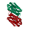 4hr0S S: Starting model for refinement C: citing same article ( |
|---|---|
| Similar structure data |
-Links
 PDBj
PDBj

- Assembly
| Deposited unit | 
| ||||||||||||
|---|---|---|---|---|---|---|---|---|---|---|---|---|---|
| 1 | 
| ||||||||||||
| Unit cell |
|
-Components
| #1: Protein | Mass: 37060.535 Da / Num. of mol.: 1 Source method: isolated from a genetically manipulated source Source: (gene. exp.) Sulfolobus acidocaldarius DSM 639 (acidophilic)Gene: Saci_1112 / Production host:  |
|---|---|
| #2: Chemical | ChemComp-MN3 /   Mass: 54.938 Da / Num. of mol.: 1 / Source method: obtained synthetically / Formula: Mn Mass: 54.938 Da / Num. of mol.: 1 / Source method: obtained synthetically / Formula: Mn |
| #3: Chemical | ChemComp-FE /   Mass: 55.845 Da / Num. of mol.: 1 / Source method: obtained synthetically / Formula: Fe Mass: 55.845 Da / Num. of mol.: 1 / Source method: obtained synthetically / Formula: Fe |
| #4: Water | ChemComp-HOH /  Mass: 18.015 Da / Num. of mol.: 53 / Source method: isolated from a natural source / Formula: H2O Mass: 18.015 Da / Num. of mol.: 53 / Source method: isolated from a natural source / Formula: H2O |
| Has ligand of interest | N |
-Experimental details
-Experiment
| Experiment | Method: X-RAY DIFFRACTION / Number of used crystals: 1 |
|---|
- Sample preparation
| Crystal | Density Matthews: 2.67 Å3/Da / Density % sol: 54.01 % |
|---|---|
| Crystal grow | Temperature: 294 K / Method: vapor diffusion, sitting drop / pH: 9 Details: 24% (w/v) polyethylene glycol 1500, 5% (v/v) formamide, 40 mM sodium propionate, 20 mM sodium cacodylate trihydrate, 40 mM bis-tris propane |
-Data collection
| Diffraction | Mean temperature: 100 K / Serial crystal experiment: N |
|---|---|
| Diffraction source | Source: SYNCHROTRON / Site: SLS  / Beamline: X06SA / Wavelength: 0.984 Å / Beamline: X06SA / Wavelength: 0.984 Å |
| Detector | Type: DECTRIS PILATUS 6M-F / Detector: PIXEL / Date: Nov 1, 2014 |
| Radiation | Protocol: SINGLE WAVELENGTH / Monochromatic (M) / Laue (L): M / Scattering type: x-ray |
| Radiation wavelength | Wavelength: 0.984 Å / Relative weight: 1 |
| Reflection | Resolution: 2.26→41.91 Å / Num. obs: 17196 / % possible obs: 91.8 % / Redundancy: 3.6 % / Biso Wilson estimate: 41.35 Å2 / CC1/2: 0.991 / CC star: 0.998 / Rmerge(I) obs: 0.1587 / Rpim(I) all: 0.0948 / Rrim(I) all: 0.1858 / Net I/σ(I): 6.67 |
| Reflection shell | Resolution: 2.26→2.341 Å / Redundancy: 3.7 % / Rmerge(I) obs: 1.442 / Mean I/σ(I) obs: 0.91 / Num. unique obs: 1739 / CC1/2: 0.398 / CC star: 0.755 / Rpim(I) all: 0.8499 / Rrim(I) all: 1.683 / % possible all: 93.53 |
- Processing
| Software |
| |||||||||||||||||||||||||||||||||||||||||||||||||
|---|---|---|---|---|---|---|---|---|---|---|---|---|---|---|---|---|---|---|---|---|---|---|---|---|---|---|---|---|---|---|---|---|---|---|---|---|---|---|---|---|---|---|---|---|---|---|---|---|---|---|
| Refinement | Method to determine structure: MOLECULAR REPLACEMENT Starting model: 4hr0 Resolution: 2.26→41.91 Å / SU ML: 0.3396 / Cross valid method: FREE R-VALUE / σ(F): 1.37 / Phase error: 27.3506 Stereochemistry target values: GeoStd + Monomer Library + CDL v1.2
| |||||||||||||||||||||||||||||||||||||||||||||||||
| Solvent computation | Shrinkage radii: 0.9 Å / VDW probe radii: 1.11 Å / Solvent model: FLAT BULK SOLVENT MODEL | |||||||||||||||||||||||||||||||||||||||||||||||||
| Displacement parameters | Biso mean: 46.27 Å2 | |||||||||||||||||||||||||||||||||||||||||||||||||
| Refinement step | Cycle: LAST / Resolution: 2.26→41.91 Å
| |||||||||||||||||||||||||||||||||||||||||||||||||
| Refine LS restraints |
| |||||||||||||||||||||||||||||||||||||||||||||||||
| LS refinement shell |
|
