 Movie
Movie Controller

[English] 日本語
 Yorodumi
- PDB-7qaa: Crystal structure of RARalpha/RXRalpha ligand binding domain hete... -
+ Open data

- Basic information
| Entry | Database: PDB / ID: 7qaa | ||||||
|---|---|---|---|---|---|---|---|
| Title | Crystal structure of RARalpha/RXRalpha ligand binding domain heterodimer in complex with BMS614 and oleic acid | ||||||
 Components Components |
| ||||||
 Keywords Keywords | GENE REGULATION / Nuclear receptor | ||||||
| Function / homology |  Function and homology information Function and homology informationTranscriptional regulation of granulopoiesis / Carnitine shuttle / Transcriptional regulation of white adipocyte differentiation / Signaling by Retinoic Acid / Sertoli cell fate commitment / positive regulation of binding / NR1H2 & NR1H3 regulate gene expression to control bile acid homeostasis / NR1H3 & NR1H2 regulate gene expression linked to cholesterol transport and efflux / SUMOylation of intracellular receptors / trachea cartilage development ...Transcriptional regulation of granulopoiesis / Carnitine shuttle / Transcriptional regulation of white adipocyte differentiation / Signaling by Retinoic Acid / Sertoli cell fate commitment / positive regulation of binding / NR1H2 & NR1H3 regulate gene expression to control bile acid homeostasis / NR1H3 & NR1H2 regulate gene expression linked to cholesterol transport and efflux / SUMOylation of intracellular receptors / trachea cartilage development / Recycling of bile acids and salts / Synthesis of bile acids and bile salts / glandular epithelial cell development / visceral serous pericardium development / Nuclear Receptor transcription pathway / Synthesis of bile acids and bile salts via 7alpha-hydroxycholesterol / Synthesis of bile acids and bile salts via 27-hydroxycholesterol / ventricular cardiac muscle cell differentiation / mesenchyme development / Endogenous sterols / chondroblast differentiation / embryonic camera-type eye development / positive regulation of translational initiation by iron / MLL4 and MLL3 complexes regulate expression of PPARG target genes in adipogenesis and hepatic steatosis / maternal placenta development / growth plate cartilage development / protein kinase B binding / positive regulation of T-helper 2 cell differentiation / prostate gland development / angiogenesis involved in coronary vascular morphogenesis / negative regulation of granulocyte differentiation / Regulation of lipid metabolism by PPARalpha / retinoic acid-responsive element binding / negative regulation of cartilage development / Cytoprotection by HMOX1 / regulation of hematopoietic progenitor cell differentiation / nuclear protein quality control by the ubiquitin-proteasome system / secretory columnal luminar epithelial cell differentiation involved in prostate glandular acinus development / positive regulation of thyroid hormone receptor signaling pathway / positive regulation of interleukin-5 production / positive regulation of interleukin-13 production / retinoic acid binding / nuclear retinoic acid receptor binding / camera-type eye development / outflow tract septum morphogenesis / cardiac muscle cell differentiation / positive regulation of vitamin D receptor signaling pathway / TGFBR3 expression / response to vitamin A / nuclear vitamin D receptor binding / apoptotic cell clearance / Signaling by Retinoic Acid / nuclear thyroid hormone receptor binding / heterocyclic compound binding / ureteric bud development / RNA polymerase II intronic transcription regulatory region sequence-specific DNA binding / regulation of myelination / regulation of branching involved in prostate gland morphogenesis / DNA-binding transcription repressor activity / DNA binding domain binding / ventricular cardiac muscle tissue morphogenesis / limb development / LBD domain binding / positive regulation of interleukin-4 production / positive regulation of lipoprotein transport / face development / nuclear steroid receptor activity / protein kinase A binding / negative regulation of type II interferon production / germ cell development / alpha-actinin binding / cellular response to estrogen stimulus / negative regulation of tumor necrosis factor production / monocyte differentiation / response to retinoic acid / cellular response to low-density lipoprotein particle stimulus / positive regulation of bone mineralization / cardiac muscle cell proliferation / retinoic acid receptor signaling pathway / heart morphogenesis / cellular response to retinoic acid / peptide binding / peroxisome proliferator activated receptor signaling pathway / cell maturation / positive regulation of cell cycle / hormone-mediated signaling pathway / response to cytokine / positive regulation of neuron differentiation / embryo implantation / negative regulation of miRNA transcription / placenta development / response to glucocorticoid / mRNA regulatory element binding translation repressor activity / transcription coregulator binding / RNA polymerase II transcription regulatory region sequence-specific DNA binding / SUMOylation of intracellular receptors / hippocampus development / neural tube closure / liver development / female pregnancy Similarity search - Function | ||||||
| Biological species |   Homo sapiens (human) Homo sapiens (human) | ||||||
| Method |  X-RAY DIFFRACTION / SYNCHROTRON / MOLECULAR REPLACEMENT / Resolution: 2.76 Å X-RAY DIFFRACTION / SYNCHROTRON / MOLECULAR REPLACEMENT / Resolution: 2.76 Å | ||||||
 Authors Authors | le Maire, A. / Vivat, V. / Guee, L. / Blanc, P. / Malosse, C. / Chamot-Rooke, J. / Germain, P. / Bourguet, w. | ||||||
| Funding support | 1items
| ||||||
 Citation Citation | Journal: J.Mol.Endocrinol. / Year: 2022 Title: Design and in vitro characterization of RXR variants as tools to investigate the biological role of endogenous rexinoids. Authors: le Maire, A. / Rey, M. / Vivat, V. / Guee, L. / Blanc, P. / Malosse, C. / Chamot-Rooke, J. / Germain, P. / Bourguet, W. | ||||||
| History |
|
- Structure visualization
| Structure viewer | Molecule: MolmilJmol/JSmol |
|---|
- Downloads & links
-Download
| PDBx/mmCIF format | 7qaa.cif.gz | 129.4 KB | Display | PDBx/mmCIF format |
|---|---|---|---|---|
| PDB format | pdb7qaa.ent.gz | 79.7 KB | Display | PDB format |
| PDBx/mmJSON format | 7qaa.json.gz | Tree view | PDBx/mmJSON format | |
| Others |  Other downloads Other downloads |
-Validation report
| Arichive directory | https://data.pdbj.org/pub/pdb/validation_reports/qa/7qaaftp://data.pdbj.org/pub/pdb/validation_reports/qa/7qaa | HTTPS FTP |
|---|
-Related structure data
| Related structure data |  7pdqC 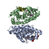 7pdtC 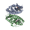 1dkfS S: Starting model for refinement C: citing same article ( |
|---|---|
| Similar structure data |
-Links
 PDBj
PDBj


- Assembly
| Deposited unit | 
| ||||||||||||
|---|---|---|---|---|---|---|---|---|---|---|---|---|---|
| 1 |
| ||||||||||||
| Unit cell |
|
-Components
| #1: Protein | Mass: 26044.152 Da / Num. of mol.: 1 Source method: isolated from a genetically manipulated source Source: (gene. exp.)  |
|---|---|
| #2: Protein | Mass: 26600.010 Da / Num. of mol.: 1 Source method: isolated from a genetically manipulated source Source: (gene. exp.) Homo sapiens (human) / Gene: RARA, NR1B1 / Production host: |
| #3: Chemical | ChemComp-OLA /   Mass: 282.461 Da / Num. of mol.: 1 / Source method: obtained synthetically / Formula: C18H34O2 / Feature type: SUBJECT OF INVESTIGATION Mass: 282.461 Da / Num. of mol.: 1 / Source method: obtained synthetically / Formula: C18H34O2 / Feature type: SUBJECT OF INVESTIGATION |
| #4: Chemical | ChemComp-BMS / 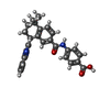  Mass: 450.528 Da / Num. of mol.: 1 / Source method: obtained synthetically / Formula: C29H26N2O3 / Feature type: SUBJECT OF INVESTIGATION Mass: 450.528 Da / Num. of mol.: 1 / Source method: obtained synthetically / Formula: C29H26N2O3 / Feature type: SUBJECT OF INVESTIGATION |
| #5: Water | ChemComp-HOH /  Mass: 18.015 Da / Num. of mol.: 121 / Source method: isolated from a natural source / Formula: H2O Mass: 18.015 Da / Num. of mol.: 121 / Source method: isolated from a natural source / Formula: H2O |
| Has ligand of interest | Y |
-Experimental details
-Experiment
| Experiment | Method: X-RAY DIFFRACTION / Number of used crystals: 1 |
|---|
- Sample preparation
| Crystal | Density Matthews: 3.87 Å3/Da / Density % sol: 68.24 % |
|---|---|
| Crystal grow | Temperature: 297 K / Method: vapor diffusion, hanging drop / Details: 23% PEG 10,000 0.1 M Hepes, pH 7.25 |
-Data collection
| Diffraction | Mean temperature: 120 K / Serial crystal experiment: N |
|---|---|
| Diffraction source | Source: SYNCHROTRON / Site: ESRF  / Beamline: ID14-1 / Wavelength: 0.918 Å / Beamline: ID14-1 / Wavelength: 0.918 Å |
| Detector | Type: MAR CCD 130 mm / Detector: CCD / Date: Dec 5, 1997 |
| Radiation | Protocol: SINGLE WAVELENGTH / Monochromatic (M) / Laue (L): M / Scattering type: x-ray |
| Radiation wavelength | Wavelength: 0.918 Å / Relative weight: 1 |
| Reflection | Resolution: 2.76→19.47 Å / Biso Wilson estimate: 47.78 Å2 |
| Reflection shell | Resolution: 2.76→2.86 Å |
- Processing
| Software |
| ||||||||||||||||||||||||||||||||||||||||||||||||||||||||
|---|---|---|---|---|---|---|---|---|---|---|---|---|---|---|---|---|---|---|---|---|---|---|---|---|---|---|---|---|---|---|---|---|---|---|---|---|---|---|---|---|---|---|---|---|---|---|---|---|---|---|---|---|---|---|---|---|---|
| Refinement | Method to determine structure: MOLECULAR REPLACEMENT Starting model: 1dkf Resolution: 2.76→19.82 Å / SU ML: 0.2487 / Cross valid method: FREE R-VALUE / σ(F): 0 / Phase error: 23.2324 Stereochemistry target values: GeoStd + Monomer Library + CDL v1.2
| ||||||||||||||||||||||||||||||||||||||||||||||||||||||||
| Solvent computation | Shrinkage radii: 0.9 Å / VDW probe radii: 1.11 Å / Solvent model: FLAT BULK SOLVENT MODEL | ||||||||||||||||||||||||||||||||||||||||||||||||||||||||
| Displacement parameters | Biso mean: 47.51 Å2 | ||||||||||||||||||||||||||||||||||||||||||||||||||||||||
| Refinement step | Cycle: LAST / Resolution: 2.76→19.82 Å
| ||||||||||||||||||||||||||||||||||||||||||||||||||||||||
| Refine LS restraints |
| ||||||||||||||||||||||||||||||||||||||||||||||||||||||||
| LS refinement shell |
|
