 Movie
Movie Controller


+ Open data

- Basic information
| Entry | Database: PDB / ID: 7q9e | ||||||
|---|---|---|---|---|---|---|---|
| Title | CYP106A1 | ||||||
 Components Components | Cytochrome P450 | ||||||
 Keywords Keywords | BIOSYNTHETIC PROTEIN / Cytochrome P450 / Steroid dehydrogenation | ||||||
| Function / homology |  Function and homology information Function and homology informationOxidoreductases; Acting on paired donors, with incorporation or reduction of molecular oxygen; With reduced flavin or flavoprotein as one donor, and incorporation of one atom of oxygen into the other donor / oxidoreductase activity, acting on paired donors, with incorporation or reduction of molecular oxygen / monooxygenase activity / iron ion binding / heme binding Similarity search - Function | ||||||
| Biological species |  Bacillus megaterium (bacteria) Bacillus megaterium (bacteria) | ||||||
| Method |  X-RAY DIFFRACTION / SYNCHROTRON / MOLECULAR REPLACEMENT / Resolution: 1.7 Å X-RAY DIFFRACTION / SYNCHROTRON / MOLECULAR REPLACEMENT / Resolution: 1.7 Å | ||||||
 Authors Authors | Carius, Y. / Hutter, M. / Kiss, F. / Bernhardt, R. / Lancaster, C.R.D. | ||||||
| Funding support |  Germany, 1items Germany, 1items
| ||||||
 Citation Citation | Journal: Febs Lett. / Year: 2022 Title: Structural comparison of the cytochrome P450 enzymes CYP106A1 and CYP106A2 provides insight into their differences in steroid conversion. Authors: Carius, Y. / Hutter, M. / Kiss, F. / Bernhardt, R. / Lancaster, C.R.D. | ||||||
| History |
|
- Structure visualization
| Structure viewer | Molecule: MolmilJmol/JSmol |
|---|
- Downloads & links
-Download
| PDBx/mmCIF format | 7q9e.cif.gz | 349.8 KB | Display | PDBx/mmCIF format |
|---|---|---|---|---|
| PDB format | pdb7q9e.ent.gz | 282.2 KB | Display | PDB format |
| PDBx/mmJSON format | 7q9e.json.gz | Tree view | PDBx/mmJSON format | |
| Others |  Other downloads Other downloads |
-Validation report
| Summary document | 7q9e_validation.pdf.gz | 2 MB | Display | wwPDB validaton report |
|---|---|---|---|---|
| Full document | 7q9e_full_validation.pdf.gz | 2.1 MB | Display | |
| Data in XML | 7q9e_validation.xml.gz | 68.3 KB | Display | |
| Data in CIF | 7q9e_validation.cif.gz | 98.8 KB | Display | |
| Arichive directory | https://data.pdbj.org/pub/pdb/validation_reports/q9/7q9eftp://data.pdbj.org/pub/pdb/validation_reports/q9/7q9e | HTTPS FTP |
-Related structure data
| Related structure data | 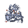 7zzlC  4yt3S S: Starting model for refinement C: citing same article ( |
|---|---|
| Similar structure data |
-Links
 PDBj
PDBj


- Assembly
| Deposited unit | 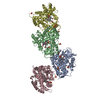
| ||||||||
|---|---|---|---|---|---|---|---|---|---|
| 1 | 
| ||||||||
| 2 | 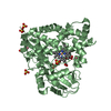
| ||||||||
| 3 | 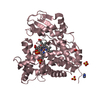
| ||||||||
| 4 | 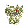
| ||||||||
| Unit cell |
|
-Components
-Protein , 1 types, 4 molecules ABCD
| #1: Protein | Mass: 48459.672 Da / Num. of mol.: 4 Source method: isolated from a genetically manipulated source Source: (gene. exp.) Bacillus megaterium (strain DSM 319) (bacteria)Strain: DSM 319 / Gene: BMD_1855 / Production host: References: UniProt: D5DF35, Oxidoreductases; Acting on paired donors, with incorporation or reduction of molecular oxygen; With reduced flavin or flavoprotein as one donor, and incorporation of one ...References: UniProt: D5DF35, Oxidoreductases; Acting on paired donors, with incorporation or reduction of molecular oxygen; With reduced flavin or flavoprotein as one donor, and incorporation of one atom of oxygen into the other donor |
|---|
-Non-polymers , 8 types, 1051 molecules 


| #2: Chemical | ChemComp-HEM /  Mass: 616.487 Da / Num. of mol.: 4 / Source method: obtained synthetically / Formula: C34H32FeN4O4 / Feature type: SUBJECT OF INVESTIGATION Mass: 616.487 Da / Num. of mol.: 4 / Source method: obtained synthetically / Formula: C34H32FeN4O4 / Feature type: SUBJECT OF INVESTIGATION#3: Chemical | ChemComp-SO4 /  Mass: 96.063 Da / Num. of mol.: 17 / Source method: obtained synthetically / Formula: SO4 Mass: 96.063 Da / Num. of mol.: 17 / Source method: obtained synthetically / Formula: SO4#4: Chemical | ChemComp-DMS / |  Mass: 78.133 Da / Num. of mol.: 1 / Source method: obtained synthetically / Formula: C2H6OS / Comment: DMSO, precipitant*YM Mass: 78.133 Da / Num. of mol.: 1 / Source method: obtained synthetically / Formula: C2H6OS / Comment: DMSO, precipitant*YM#5: Chemical |  Mass: 106.120 Da / Num. of mol.: 2 / Source method: obtained synthetically / Formula: C4H10O3 Mass: 106.120 Da / Num. of mol.: 2 / Source method: obtained synthetically / Formula: C4H10O3#6: Chemical |  Mass: 58.933 Da / Num. of mol.: 3 / Source method: obtained synthetically / Formula: Co Mass: 58.933 Da / Num. of mol.: 3 / Source method: obtained synthetically / Formula: Co#7: Chemical | ChemComp-IMD / |  Mass: 69.085 Da / Num. of mol.: 1 / Source method: obtained synthetically / Formula: C3H5N2 Mass: 69.085 Da / Num. of mol.: 1 / Source method: obtained synthetically / Formula: C3H5N2#8: Chemical | ChemComp-PEO / |  Mass: 34.015 Da / Num. of mol.: 1 / Source method: obtained synthetically / Formula: H2O2 Mass: 34.015 Da / Num. of mol.: 1 / Source method: obtained synthetically / Formula: H2O2#9: Water | ChemComp-HOH / | Mass: 18.015 Da / Num. of mol.: 1022 / Source method: isolated from a natural source / Formula: H2O |
|---|
-Details
| Has ligand of interest | Y |
|---|
-Experimental details
-Experiment
| Experiment | Method: X-RAY DIFFRACTION / Number of used crystals: 1 |
|---|
- Sample preparation
| Crystal | Density Matthews: 2.3 Å3/Da / Density % sol: 46.53 % |
|---|---|
| Crystal grow | Temperature: 291 K / Method: vapor diffusion, sitting drop / Details: PEG 3350, lithium sulphate |
-Data collection
| Diffraction | Mean temperature: 100 K / Serial crystal experiment: N |
|---|---|
| Diffraction source | Source: SYNCHROTRON / Site: ESRF  / Beamline: ID23-2 / Wavelength: 0.97916 Å / Beamline: ID23-2 / Wavelength: 0.97916 Å |
| Detector | Type: DECTRIS PILATUS 6M / Detector: PIXEL / Date: Sep 26, 2014 |
| Radiation | Protocol: SINGLE WAVELENGTH / Monochromatic (M) / Laue (L): M / Scattering type: x-ray |
| Radiation wavelength | Wavelength: 0.97916 Å / Relative weight: 1 |
| Reflection | Resolution: 1.7→25.92 Å / Num. obs: 168988 / % possible obs: 88.9 % / Redundancy: 3.3 % / Biso Wilson estimate: 18.6 Å2 / CC1/2: 0.996 / Rmerge(I) obs: 0.066 / Rpim(I) all: 0.043 / Rrim(I) all: 0.079 / Net I/σ(I): 7.05 |
| Reflection shell | Resolution: 1.7→1.79 Å / Rmerge(I) obs: 0.307 / Mean I/σ(I) obs: 2.11 / Num. unique obs: 23968 / CC1/2: 0.92 / Rpim(I) all: 0.199 / Rrim(I) all: 0.367 |
- Processing
| Software |
| ||||||||||||||||||||||||||||||||||||||||||||||||||||||||||||
|---|---|---|---|---|---|---|---|---|---|---|---|---|---|---|---|---|---|---|---|---|---|---|---|---|---|---|---|---|---|---|---|---|---|---|---|---|---|---|---|---|---|---|---|---|---|---|---|---|---|---|---|---|---|---|---|---|---|---|---|---|---|
| Refinement | Method to determine structure: MOLECULAR REPLACEMENT Starting model: 4YT3 Resolution: 1.7→25.82 Å / Cor.coef. Fo:Fc: 0.963 / Cor.coef. Fo:Fc free: 0.945 / SU B: 2.118 / SU ML: 0.07 / Cross valid method: THROUGHOUT / σ(F): 0 / ESU R: 0.111 / ESU R Free: 0.109 / Stereochemistry target values: MAXIMUM LIKELIHOOD Details: HYDROGENS HAVE BEEN ADDED IN THE RIDING POSITIONS U VALUES : REFINED INDIVIDUALLY
| ||||||||||||||||||||||||||||||||||||||||||||||||||||||||||||
| Solvent computation | Ion probe radii: 0.8 Å / Shrinkage radii: 0.8 Å / VDW probe radii: 1.2 Å / Solvent model: MASK | ||||||||||||||||||||||||||||||||||||||||||||||||||||||||||||
| Displacement parameters | Biso max: 89.25 Å2 / Biso mean: 22.634 Å2 / Biso min: 7.36 Å2
| ||||||||||||||||||||||||||||||||||||||||||||||||||||||||||||
| Refinement step | Cycle: final / Resolution: 1.7→25.82 Å
| ||||||||||||||||||||||||||||||||||||||||||||||||||||||||||||
| Refine LS restraints |
| ||||||||||||||||||||||||||||||||||||||||||||||||||||||||||||
| LS refinement shell | Resolution: 1.7→1.744 Å / Rfactor Rfree error: 0 / Total num. of bins used: 20
|
